. 2016 Jul 12;55(27):3834-46.
doi: 10.1021/acs.biochem.6b00203. Epub 2016 Jun 27.

Molecular Dynamics Study of Twister Ribozyme: Role of Mg(2+) Ions and the Hydrogen-Bonding Network in the Active Site

Affiliations

Molecular Dynamics Study of Twister Ribozyme: Role of Mg(2+) Ions and the Hydrogen-Bonding Network in the Active Site

Melek N Ucisik et al. Biochemistry. .

Abstract

The recently discovered twister ribozyme is thought to utilize general acid-base catalysis in its self-cleavage mechanism, but the roles of nucleobases and metal ions in the mechanism are unclear. Herein, molecular dynamics simulations of the env22 twister ribozyme are performed to elucidate the structural and equilibrium dynamical properties, as well as to examine the role of Mg(2+) ions and possible candidates for the general base and acid in the self-cleavage mechanism. The active site region and the ends of the pseudoknots were found to be less mobile than other regions of the ribozyme, most likely providing structural stability and possibly facilitating catalysis. A purported catalytic Mg(2+) ion and the closest neighboring Mg(2+) ion remained chelated and relatively immobile throughout the microsecond trajectories, although removal of these Mg(2+) ions did not lead to any significant changes in the structure or equilibrium motions of the ribozyme on the microsecond time scale. In addition, a third metal ion, a Na(+) ion remained close to A1(O5'), the leaving group atom, during the majority of the microsecond trajectories, suggesting that it might stabilize the negative charge on A1(O5') during self-cleavage. The locations of these cations and their interactions with key nucleotides in the active site suggest that they may be catalytically relevant. The P1 stem is partially melted at its top and bottom in the crystal structure and further unwinds in the trajectories. The simulations also revealed an interconnected network comprised of hydrogen-bonding and π-stacking interactions that create a relatively rigid network around the self-cleavage site. The nucleotides involved in this network are among the highly conserved nucleotides in twister ribozymes, suggesting that this interaction network may be important to structure and function.


Conflict of interest statement

The authors declare no competing financial interest.

Figures

Figure 1
Figure 1
(A) Structure of the env22 twister ribozyme (PDB entry 4RGE). The U–1-A1 self-cleavage site is depicted as sticks with nitrogen and oxygen atoms colored blue and red, respectively, and the crystallographic Mg2+ ions are depicted as pink spheres. The active site Mg2+ ion, Mg101, is depicted as a cyan sphere, and a nearby Mg2+ ion, Mg109, is slightly visible in pink behind the ribozyme upward to the right of Mg101. Pseudoknots T1 and T2 are colored magenta and orange, respectively, and the P1 stem is colored light green. (B) Schematic depiction of the secondary structure of the same ribozyme. The self-cleavage site is marked with a black triangle. The pseudoknots and P1 stem are depicted using the same color scheme as in part A. Pseudoknots T1 and T2 are labeled, as well. The interactions of the residues denoted as P1 are drawn according to PDB entry 4RGE. (C) Schematic depiction of the site-specific phosphodiester bond cleavage in the env22 twister ribozyme at the phosphate linking nucleotides U–1 and A1.
Figure 2
Figure 2
Time evolution of the RMSDs for the independent MD trajectories. The black, red, and green lines represent the data from three independent trajectories containing all crystallographic Mg2+ ions. The blue line represents the data from a trajectory with Mg109 replaced by Na+, the purple line the data from a trajectory with Mg101 replaced by Na+, and the cyan line the data from a trajectory with no Mg2+ ions.
Figure 3
Figure 3
(A) RMSFs of the P atoms for the 19-mer substrate and the 37-mer ribozyme strands of the env22 twister ribozyme. The break at residue 15 arises because it is the 5′-terminal nucleotide of the 37-mer. The black, red, and green lines represent the data from three independent trajectories containing all crystallographic Mg2+ ions. The blue line represents the data from a trajectory with Mg109 replaced by Na+, the purple line the data from a trajectory with Mg101 replaced by Na+, and the cyan line the data from a trajectory with no Mg2+ ions. (B) Depiction of the regions with low RMSFs. The cyan nucleotides are U–1 and A1, which constitute the self-cleavage site. The green region contains C9 and A10, while the blue portions feature nucleotides 25–47. Note that active site residues −1, 1, 25, 27, 29, 30, and 40–43 are associated with a lower RMSF.
Figure 4
Figure 4
(A) Time evolution and (B) histograms for the hydrogen bonds formed between the nucleotides. The black, red, and green lines represent the data from three independent trajectories containing all 10 crystallographic Mg2+ ions. The blue line represents the data from a trajectory with Mg109 replaced by Na+, the purple line the data from a trajectory with Mg101 replaced by Na+, and the cyan line the data from a trajectory with no Mg2+ ions.
Figure 5
Figure 5
Depiction of the active site of the env22 twister ribozyme for a representative configuration from the “full run 1” trajectory. G43 (in ball and stick representation) is hypothesized to participate in the self-cleavage mechanism by stabilizing the negatively charged transition state through the hydrogen bonds it forms with the pro-RP oxygen of the scissile phosphate, shown as black dashed lines. Mg101 is observed to coordinate to the pro-SP oxygen of the scissile phosphate and thus is a candidate to actively contribute to the self-cleavage mechanism. It also coordinates to the pro-RP oxygen of A2 and O4 of U3, as well as three water molecules (cyan dashed lines). A1 displays a syn conformation at all times.
Figure 6
Figure 6
(A) Radial distribution functions for Mg2+ and Na+ ions relative to the pro-SP (OP1) oxygen of A1 and the pro-RP (OP2) oxygen of A2 in the region of Mg101. The radial distribution functions for Mg2+ (black and red curves, “full run 1”) form pronounced peaks at ∼2.0 Å, with a second peak indicating that another Mg2+ is located at a distance of ∼4.2 Å for A2(OP2). The results for “full run 1” are representative of those for “full runs 1–3”. The radial distribution functions for Na+ (blue and cyan curves, “Na+ at Mg101”) form smaller peaks at ∼2.1 Å because of the higher density of Na+ in the simulation cell, given that the ion density occurs in the denominator of the standard definition of the radial distribution function. See the Supporting Information for the standard definitions of the radial distribution function and running coordination number. (B) Running coordination numbers for Mg2+ or Na+ relative to the pro-SP oxygen (OP1) of A1 and the pro-RP oxygen (OP2) of A2 in the region of Mg101. A shared Mg2+ or a shared Na+ is coordinated to both oxygen atoms at ∼2.0 Å for the “full run 1” or “Na+ at Mg101” trajectory, respectively. The coordination number for Na+ to A1(OP1) is slightly greater than unity at ∼2.0 Å, indicating that another Na+ samples this spherical shell in some configurations. The second step for the “full run 1” trajectory indicates that another Mg2+ is located at a distance of ∼4.2 Å from A2(OP2). Note that there is no practicing consensus in the RNA literature about the assignment of OP1. Thus, we used the terminology in ref (19), which is to assign OP1 as pro-SP.
Figure 7
Figure 7
Relative positioning of A1, A29, and A30 for a representative configuration from (A) the “full run 1” trajectory, in the presence of Mg2+ ions, illustrating a “T-shaped” stacking interaction between A29 and A1 and (B) the “no Mg2+” trajectory, in the absence of Mg2+ ions, illustrating a π–π stacking interaction between A1 and A30.
Figure 8
Figure 8
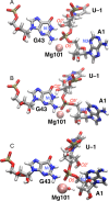
Evolution of active site nucleotides U–1, A1, and G43 in the MD simulations based on the 4RGE crystal structure. (A) Crystal structure geometry after substitution of the hydroxyl group at U–1(C2′) and a local optimization of its position. (B) Structure following geometry optimization of the entire ribozyme. (C) Configuration obtained after MD for 2 μs for the “full run 1” trajectory. The G43(N1)–U–1(O2′) distance is larger in part C than in parts A and B. A1(O5′) is hydrogen bonded to a Mg2+-bound water molecule in part B but then rotates to point toward A1(N3) in part C; the A1(O5′)–A1(N3) distance is smaller in part C (∼4.4 Å) than in part B (∼5.4 Å). The U–1(O2′)–A1(P)–A1(O5′) angle is more linear in part A than in parts B and C. The 5DUN crystal structure is similar to the configuration in part C.
Figure 9
Figure 9
Active site region for a representative configuration obtained from the “full run 1” trajectory. Possible general base candidates for the deprotonation of U–1(O2′), which is marked with a red label, and possible general acid candidates for the protonation of A1(O5′), which is also marked with a red label, are annotated. Mg101 is shown as a pink sphere.
Figure 10
Figure 10
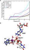
(A) Running coordination numbers for Na+ relative to A1(O5′). All trajectories except for “full run 3” exhibited a Na+ near this O5′. (B) Representative configuration from the “full run 1” trajectory depicting the Na+ ion as a purple sphere between A1(N3) and A1(O5′) located directly above the ribose of A1. This Na+ ion is coordinated to A1(N3), A1(O4′), A1(O5′), U−2(O2′), and two water molecules, although the coordination to U−2(O2′) is not shown here.
Figure 11
Figure 11
Set of nucleotides lining the catalytic pocket for a representative configuration from the “full run 1” trajectory. The backbone ribose and phosphate of each nucleotide are omitted for the sake of simplicity. U–1 and A1 are displayed as thicker sticks. The water-mediated hydrogen bonding among C41(N4), A42(N6), and U–1(O2′) is shown with black dotted lines. The catalytic Mg101 is shown as a pink sphere.

Similar articles

Cited by

References

    1. Benner S. A.; Ellington A. D.; Tauer A. (1989) Modern metabolism as a palimpsest of the RNA world. Proc. Natl. Acad. Sci. U. S. A. 86, 7054–7058. 10.1073/pnas.86.18.7054. - DOI - PMC - PubMed
    1. Orgel L. E. (2004) Prebiotic chemistry and the origin of the RNA world. Crit. Rev. Biochem. Mol. Biol. 39, 99–123. 10.1080/10409230490460765. - DOI - PubMed
    1. Cech T. R. (2012) The RNA worlds in context. Cold Spring Harbor Perspect. Biol. 4, a006742.10.1101/cshperspect.a006742. - DOI - PMC - PubMed
    1. Webb C. H. T.; Riccitelli N. J.; Ruminski D. J.; Luptak A. (2009) Widespread occurrence of self-cleaving ribozymes. Science 326, 953–953. 10.1126/science.1178084. - DOI - PMC - PubMed
    1. Roth A.; Weinberg Z.; Chen A. G. Y.; Kim P. B.; Ames T. D.; Breaker R. R. (2013) A widespread self-cleaving ribozyme class is revealed by bioinformatics. Nat. Chem. Biol. 10, 56–60. 10.1038/nchembio.1386. - DOI - PMC - PubMed

Publication types