. 2016 Aug 1;55(32):9411-5.
doi: 10.1002/anie.201603178. Epub 2016 Jun 13.

GM1 Ganglioside Inhibits β-Amyloid Oligomerization Induced by Sphingomyelin

Affiliations

GM1 Ganglioside Inhibits β-Amyloid Oligomerization Induced by Sphingomyelin

Mariana Amaro et al. Angew Chem Int Ed Engl. .

Abstract

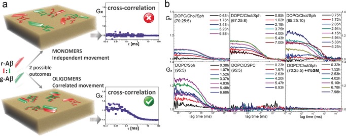
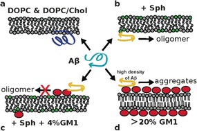
β-Amyloid (Aβ) oligomers are neurotoxic and implicated in Alzheimer's disease. Neuronal plasma membranes may mediate formation of Aβ oligomers in vivo. Membrane components sphingomyelin and GM1 have been shown to promote aggregation of Aβ; however, these studies were performed under extreme, non-physiological conditions. We demonstrate that physiological levels of GM1 , organized in nanodomains do not seed oligomerization of Aβ40 monomers. We show that sphingomyelin triggers oligomerization of Aβ40 and that GM1 is counteractive thus preventing oligomerization. We propose a molecular explanation that is supported by all-atom molecular dynamics simulations. The preventive role of GM1 in the oligomerization of Aβ40 suggests that decreasing levels of GM1 in the brain, for example, due to aging, could reduce protection against Aβ oligomerization and contribute to the onset of Alzheimer's disease.

Keywords: Alzheimer's disease; amyloid beta-peptides; diffusion coefficients; fluorescence spectroscopy; neuroprotectives.


Figures

Figure 1
Figure 1
a) Lateral diffusion coefficients, D 2D, of membrane‐bound Aβ (g‐Aβ) and lipid tracers (DiD and g‐GM1) in DOPC/Chol/Sph GUVs containing 0 or 4 % added GM1. A 2D component model describes well the diffusion of Aβ and lipid tracers. D 2D of Aβ decreases with time (green arrow symbols; see next panel) in DOPC/Chol/Sph bilayers, indicating oligomerization of Aβ. In the quaternary compositions [(DOPC/Chol/Sph)+4 %GM1)] no changes of Aβ diffusion are observed. b) Time evolution of D 2D of membrane‐bound Aβ (t“; 0 h, addition of Aβ monomers). DOPC/Chol/Sph compositions: circles (70:25.5); triangles (67:25:8); squares (65:25:10). Each point is the weighted average of D 2D results obtained from at least five independent two‐color Z‐FCS measurements (each composed of 15–20 scans). Error bars are the standard deviation within the sample of D 2D results obtained for each composition. Where D 2D of Aβ varies with time, error bars for Aβ values are the standard deviation obtained from the fitting procedure of 15 scans obtained via Z‐FCS.
Figure 2
Figure 2
a) Illustration of cross‐correlation experiments using a 1:1 mixture of r‐ and g‐Aβ monomers. Top: Membrane‐bound r‐ and g‐Aβ monomers diffuse independently; thus red and green signal fluctuations are not correlated, resulting in null cross‐correlation function (Gx), that is, Gx=1. Bottom: Formation of Aβ oligomers implies co‐diffusion of peptides. If oligomers are formed of both r‐ and g‐Aβ, red and green signal fluctuations become correlated, resulting in positive cross‐correlation, that is, Gx>1. b) Summary of results. Top: DOPC/Chol/Sph membranes show positive cross‐correlation. Bottom left: DOPC/Sph bilayers show positive cross‐correlation, example DOPC/Sph (95:5). Bottom middle: DOPC/DSPC membranes show no cross‐correlation, example DOPC/DSPC (95:5). Bottom right: DOPC/Chol/Sph+GM1 bilayers show no cross‐correlation, example [DOPC/Chol/Sph (70:25:5)+4 %GM1].
Figure 3
Figure 3
Proposed model. a) Aβ does not oligomerize when bound to DOPC and DOPC/Chol bilayers. The C‐terminus of Aβ has no secondary structure (dark blue representation). b) Oligomerization of Aβ occurs in the presence of Sph, for both DOPC/Sph and DOPC/Chol/Sph membranes. The C‐terminus of monomeric Aβ acquires β‐sheet structure (yellow representation) in bilayers containing Sph. c) Binding of Aβ to the headgroup of GM1 (red ellipses) sequesters the peptide and prevents it from forming oligomers. d) High density of both GM1 and Aβ facilitates aggregation of the peptide and fibril formation (from literature),5 which can be explained by generic surface effects.18

References

    1. None
    1. Bucciantini M., Giannoni E., Chiti F., Baroni F., Formigli L., Zurdo J., Taddei N., Ramponi G., Dobson C. M., Stefani M., Nature 2002, 416, 507–511; - PubMed
    1. Shankar G. M., Li S., Mehta T. H., Garcia-munoz A., Nina E., Smith I., Brett F. M., Farrell M. A., Rowan M. J., Lemere C. A. et al., Nat. Med. 2008, 1 4, 837–842. - PMC - PubMed
    1. None
    1. Amaro M., Birch D. J. S., Rolinski O. J., Phys. Chem. Chem. Phys. 2011, 13, 6434–6441; - PubMed

Publication types

MeSH terms

LinkOut - more resources