. 2016 Jul;18(7):765-76.
doi: 10.1038/ncb3372. Epub 2016 Jun 13.

Unconventional secretion of misfolded proteins promotes adaptation to proteasome dysfunction in mammalian cells

Affiliations

Unconventional secretion of misfolded proteins promotes adaptation to proteasome dysfunction in mammalian cells

Jin-Gu Lee et al. Nat Cell Biol. 2016 Jul.

Abstract

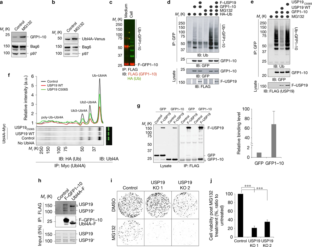
To safeguard proteomic integrity, cells rely on the proteasome to degrade aberrant polypeptides, but it is unclear how cells remove defective proteins that have escaped degradation owing to proteasome insufficiency or dysfunction. Here we report a pathway termed misfolding-associated protein secretion, which uses the endoplasmic reticulum (ER)-associated deubiquitylase USP19 to preferentially export aberrant cytosolic proteins. Intriguingly, the catalytic domain of USP19 possesses an unprecedented chaperone activity, allowing recruitment of misfolded proteins to the ER surface for deubiquitylation. Deubiquitylated cargos are encapsulated into ER-associated late endosomes and secreted to the cell exterior. USP19-deficient cells cannot efficiently secrete unwanted proteins, and grow more slowly than wild-type cells following exposure to a proteasome inhibitor. Together, our findings delineate a protein quality control (PQC) pathway that, unlike degradation-based PQC mechanisms, promotes protein homeostasis by exporting misfolded proteins through an unconventional protein secretion process.


Conflict of interest statement

COMPETING FINANCIAL INTERESTS

The authors declare no competing financial interests.

Figures

Figure 1
Figure 1
USP19 promotes secretion of a cytosolic protein. (a) The domain architecture of USP19. TM, transmembrane; UBL, ubiquitin-like; ZnF, zinc finger; CS, CHORD-containing proteins and SGT1; USP, ubiquitin-specific protease. (b) Overexpression of USP19 induces GFP secretion from HEK293T cells. Cells (0.8×10⁵) in a 12-well plate were transfected with 250ng pEGFP together with 250ng pcDNA3 or FLAG–USP19-encoding plasmid. At 24h post-transfection, the medium was replaced with fresh medium. Cells were incubated for 16h. The conditioned media and cell lysates were collected and directly analysed by immunoblotting (IB) (lanes 1 and 2). A fraction of the media was subjected to immunoprecipitation (IP) by GFP antibodies to enrich GFP (lanes 3 and 4). Asterisk indicates a nonspecific IgG band. Left panels show two different exposures (long and short). (c) USP19 but not USP7 induces GFP secretion from HEK293T cells. Conditioned media (16h) and lysates prepared from cells co-transfected with GFP together with the indicated constructs were analysed by immunoblotting. (d) Overexpression of WT USP19 or USP19C506S (the catalytically inactive mutant) does not affect PM permeability. HEK293T cells transfected with the indicated plasmids for 48h were stained with trypan blue and counted (mean ± s.e.m., n=3 independent experiments). (e) USP19 induces GFP secretion in a dose-dependent manner in HEK293T cells. The arrowhead indicates endogenous USP19. (f) WT USP19 but not the catalytically inactive mutant (C506S) induces GFP secretion from HeLa cells. (g) GFP secretion from HEK293T cells requires both the catalytic activity and the TM domain of USP19. HEK293T cells transfected as indicated were analysed as in c. (h) GFP secretion is not inhibited by brefeldin A (BFA). Media collected at the indicated time points from HEK293T cells stably expressing GFP were analysed by immunoblotting. A fraction of the samples was subjected to immunoprecipitation with GFP antibodies before immunoblotting. Where indicated, dimethylsulfoxide (DMSO; control) or BFA (10μgml−1) was added at the beginning of the chase. Asterisk, IgG band. Statistics source data for d can be found in Supplementary Table 2. Unprocessed original scans of blots are shown in Supplementary Fig. 8.
Figure 2
Figure 2
USP19 preferentially targets misfolded cytosolic proteins for secretion. (a) Secreted GFP molecules are mostly unfolded. HEK293T cells (0.5×10⁶) were transfected with 1μg GFP together with 1μg USP19. Conditioned medium collected between 36 and 52h post-transfection and cell lysate were examined for green fluorescence. Shown is GFP intensity normalized by protein level determined by immunoblotting (Supplementary Fig. 1a). (b) USP19-induced secretion requires its deubiquitylating activity and the TM domain. Conditioned media (16h) from HEK293T cells transfected with the indicated plasmids were analysed together with lysates by immunoblotting. (c) Secretion of GFP1–10 is affected by its folding state. Quantification of four independent experiments represented by Supplementary Fig. 1c. The level of GFP1–10 secretion was normalized by the GFP1–10 level in lysate (mean ± s.e.m., n = 4 independent experiments, ∗∗P < 0.01 as determined by paired Student’s t-test). (d) Kinetic analysis of GFP1–10 secretion using HEK293T cells co-transfected with GFP1–10 and the indicated plasmids. Media collected at the indicated time points (top panel) and lysates prepared at the end of the chase (bottom panels) were analysed by immunoblotting. (e) Overexpression of ER-tethered USP7 does not promote GFP1–10 secretion. The same as in b, except that the indicated deubiquitylases were transfected. (f) USP19 does not induce secretion of endogenous cytosolic proteins. Conditioned media and lysates from HEK293T cells transfected with control or USP19 were analysed by immunoblotting. (g) USP19 promotes secretion of overexpressed Ubl4A. HEK293T cells co-transfected with Ubl4A–FLAG together with the indicated USP19 variants were analysed as in b. (h) USP19 promotes α-synuclein secretion. As in g, except that Ubl4A–FLAG was replaced by α-synuclein–FLAG in transfection. (i) USP19 promotes the secretion of Parkinson’s disease-associated α-synuclein mutants. The graph shows the relative secretion efficiency (mean ± s.e.m., n = 3 independent experiments). (j) USP19 does not promote Tau secretion. Note that Tau was not detected in media in any of the tested conditions. Statistics source data for c,i can be found in Supplementary Table 2. Unprocessed original scans of blots are shown in Supplementary Fig. 8.
Figure 3
Figure 3
Endogenous USP19 is required for MAPS. (a) Knockdown of endogenous USP19 reduces GFP1–10 secretion. HEK293T cells were transfected with GFP1–10 together with the indicated siRNAs. The conditioned media (16h) and lysates were analysed by immunoblotting. (b) Quantification of GFP1–10 secretion from HEK293T cells transfected with control and USP19 siRNA-2 (mean ± s.e.m., n = 4 independent experiments, ∗∗P < 0.01). (c) The design of the CRISPR guide RNAs that target the USP19 gene. The predicted nicking sites are labelled. (d) USP19 deficiency reduces GFP1–10 secretion. GFP1–10 secretion was analysed using a control CRISPR clone and two USP19 knockout (KO) clones as well as the parental HEK293T cells. Asterisk, a nonspecific band. (e) Quantification of GFP1–10 secretion from control and USP19-null cells (mean ± s.e.m., n = 4 independent experiments, ∗∗∗P< 0.001, ∗∗P<0.01). (f) Overexpression of USP19 rescues GFP1–10 secretion in USP19-null cells. (g,h) Knockout of USP19 impairs the secretion of overexpressed Ubl4A–FLAG. (g) The same as in d, except that control and USP19-null cells were transfected with Ubl4A–FLAG. (h) Quantification of the experiments represented in g (mean ± s.e.m., n = 3 independent experiments, ∗∗P<0.01). (i,j) Knockout of USP19 reduces the secretion of overexpressed α-synuclein–FLAG (mean ± s.e.m., n = 3 independent experiments, ∗∗∗P <0.001). Statistics source data for b,e,h,j can be found in Supplementary Table 2. Unprocessed original scans of blots are shown in Supplementary Fig. 8.
Figure 4
Figure 4
USP19 interacts with MAPS cargos and alters their ubiquitylation state. (a,b) Both GFP1–10 and Ubl4A are proteasome substrates. (a) HEK293T cells transfected with GFP1–10 were treated with MG132 (10 μM) for 16h. Lysates were subjected to immunoblot analysis. (b) The same as in a, except that cells transfected with Ubl4A–Venus were analysed. (c) GFP1–10 is ubiquitylated in cells, but secreted GFP1–10 is mostly unmodified. GFP1–10 was immunoprecipitated under denaturing conditions from lysates or conditioned media using HEK293T cells co-transfected with GFP1–10 and haemagglutinin (HA)-tagged ubiquitin. Asterisks, nonspecific bands. (d) USP19 promotes deubiquitylation of GFP1–10 in cells. HEK293T cells transfected with the indicated constructs were treated with DMSO as a control or MG132 (20 μM) for 4h. GFP1–10 immunoprecipitated from cell extracts was analysed by immunoblotting (top panels). A fraction of the lysates was analysed directly by immunoblotting (bottom panels). (e) WT USP19 but not USP19C506S reduced ubiquitylated GFP1–10. The same as in d, except that cells were transfected and treated as indicated. (f) USP19 promotes deubiquitylation of Ubl4A–FLAG. Cells transfected with the indicated constructs were treated with MG132 (20 μM) for 4h. Ubl4A–FLAG was immunoprecipitated and analysed by immunoblotting. The graph shows the intensity of ubiquitylated Ubl4A bands quantified from the gel below. (g) USP19 binds GFP1–10 more strongly than GFP. Co-immunoprecipitation using cells co-transfected with either control or FLAG–USP19 together with the indicated MAPS cargos. Asterisk, IgG bands. The graph shows the quantification result (mean ± s.e.m., n = 3 independent experiments). (h) Endogenous USP19 interacts with FLAG–GFP1–10 and Ubl4A–FLAG. Cells transfected with the indicated constructs were treated with formaldehyde. Cell lysates were subjected to immunoprecipitation by FLAG antibodies. Asterisk, nonspecific band; USP19*, a truncated USP19 product. (i,j) USP19-null cells have a growth defect after treatment with a proteasome inhibitor. (i) Control and USP19 knockout cells treated with MG132 (5 μM, 15h) or DMSO were incubated in inhibitor-free medium for 14 days and stained. (j) Quantification of relative cell viability of MG132-treated cells (mean ± s.e.m., n = 3 independent experiments). ∗∗∗P < 0.001. Statistics source data for g,j can be found in Supplementary Table 2. Unprocessed original scans of blots are shown in Supplementary Fig. 8.
Figure 5
Figure 5
USP19 has a chaperone activity. (a) Purified FLAG-tagged WT USP19 and the USP19C506S mutant. (b) An in vitro deubiquitylation assay using ubiquitin-AFC as the substrate confirms the activity of WT USP19. (c) USP19 inhibits luciferase aggregation in vitro. Luciferase incubated with an increased concentration of either WT USP19 or USP19C506S at 42 °C (15min) was subjected to centrifugation. The resulting soluble fractions and a fraction of the samples not exposed to the heat treatment were analysed by immunoblotting with FLAG (USP19) and luciferase antibodies. The numbers indicate band intensity. (d) USP19 preferentially recognizes unfolded luciferase. Luciferase incubated with WT USP19 either on ice or at 42 °C was subjected to centrifugation. The resulting soluble fractions were used for immunoprecipitation by FLAG beads. (e) The N-terminal CS domains are dispensable for recognition of unfolded luciferase. The same as in d, except that all samples were heat-treated and that purified USP19 Δ1–493 was tested together with WT USP19. (f,g) The USP19 N-terminal domain is dispensable for MAPS. Where indicated, different amounts of USP19 WT and USP19 Δ1–493 plasmids were transfected together with either GFP1–10 (f) or Ubl4A–FLAG (g). The numbers show band intensity. Unprocessed original scans of blots are shown in Supplementary Fig. 8.
Figure 6
Figure 6
USP19 recruits misfolded proteins to the ER and ER-associated late endosomes. (a) USP19 preferentially recruits GFP1–10 to membranes. The cytosol and membrane fractions from HEK293T cells co-transfected with the indicated constructs were analysed by immunoblotting. (b) Knockout of USP19 reduces membrane-associated GFP1–10. Control and USP19 CRISPR (KO) cells were transfected with GFP1–10. After fractionation, GFP1–10 in the membrane and cytosol fractions was analysed by immunoblotting. The graph shows the level of membrane-associated GFP1–10 relative to cytosolic GFP1–10 (mean ± s.e.m., n = 3 independent experiments, ∗∗P < 0.01). (c) USP19 recruits GFP1–10 to the ER membrane. COS7 cells co-transfected with GFP1–10 and FLAG-USP19 were permeabilized, stained with GFP and FLAG antibodies and analysed by confocal microscopy. Bottom panels show the outline of GFP1–10 and USP19 signal in the outlined area. Scale bar, 5 μm. (d,e) A photobleaching-based assay reveals vesicles containing MAPS cargos. (d) A schematic illustration of the photobleaching experiment shown in e. The marked area in d and e was photobleached with a 568nm laser to remove cytosolic and ER-associated mCh–GFP1–10 background. Arrows indicate a few examples of vesicles revealed after photobleaching. Scale bar, 5 μm. (f) MAPS vesicles were labelled with Rab9. Shown are two frames from a live-cell imaging experiment using cells transfected with mCe–USP19, mCi–Rab9, and mCh–GFP1–10 after photobleaching. Panels 4–6 are enlarged views of the indicated area in panel 1 after 20s. (g) Structure-illuminated microscopy analysis of MAPS vesicles. Cells transfected with mCe–USP19, Ubl4A–Venus and mCh–Rab9 were permeabilized, fixed and imaged. Note that Ubl4A–Venus is detected on the membranes of intraluminal vesicles, but not in the lumen of these vesicles. (h) Transmission electron microscopy analyses of MAPS vesicles. COS7 cells expressing FLAG-tagged GFP1–10 were permeabilized, fixed, and stained with anti-FLAG antibodies and immunogold-labelled secondary antibodies. Blue arrows show examples of luminal GFP1–10 signals. Arrowheads show PM-associated GFP1–10. The inset shows an example of GFP1–10 association with the limiting membrane on the luminal side (yellow arrows). Statistics source data for b can be found in Supplementary Table 2. Unprocessed original scans of blots are shown in Supplementary Fig. 8.
Figure 7
Figure 7
Secretion of misfolded proteins does not involve autophagosomes or exosomes. (a) Two established UPS pathways that involve an intracellular vesicle carrier. (b) GFP1–10 is not secreted by exosomes. Conditioned media (16h) collected from cells transfected with GFP1–10 together with the indicated USP19 variants were subjected to differential centrifugation and immunoblot analysis. S, supernatant; P, pellet. The faint bands in the outlined areas are caused by serum proteins crossreacting with the antibodies. (c,d) Most MAPS vesicles are not autophagosomes. COS7 cells transfected with mCh–GFP1–10 and mCe–USP19 were photobleached, methanol-fixed, and stained with LC3 antibodies. Arrowheads in c,d show examples of MAPS vesicles not labelled by LC3 antibodies. Scale bar, 5 μm. (d) A 3D reconstructed image after z-section confocal analysis. (e) Secretion of Ubl4A–FLAG is not affected by starvation. Cells transfected with the indicated plasmids were either incubated in complete medium or an EBSS starvation medium. Media collected at the indicated time points and lysates prepared at the end of the chase were analysed by immunoblotting. (f,g) GFP1–10 secretion does not require GRASPs. (f) Secretion of GFP1–10 was analysed in cells transfected with GFP1–10 and FLAG–USP19 together with the indicated siRNAs. Knockdown of GRASP65 was confirmed by immunoblotting. (h) The same as in g, except that GRASP55 and Tsg101 were included and that gene knockdown was confirmed by quantitative PCR with reverse transcription (indicated in green labels). Unprocessed original scans of blots are shown in Supplementary Fig. 8.
Figure 8
Figure 8
Secretion of misfolded proteins through late endosomes. (a–c) The number of MAPS vesicles in cells correlates with the secretion efficiency. (a) Representative cells transfected with mCherry or mCh–GFP1–10 before and after photobleaching. (b) Representative cells transfected with mCh–GFP1–10 together with either mCi–USP19 WT or the mCi–USP19C506S (CS) mutant. (c) Quantification of MAPS vesicles in photobleached COS7 cells that were transfected as indicated. (d) Confocal analysis of v-SNAREs in live cells transfected with mCh–Rab9 and the indicated EGFP-tagged v-SNAREs. (e) Expression of late endosome-localized v-SNAREs promotes GFP1–10 secretion. Secretion of GFP1–10 was analysed in cells transfected with GFP1–10 and the indicated EGFP–v-SNAREs. (f) The graph shows the quantification result from experiments represented in e (mean ± s.e.m., n=3 independent experiments, P <0.05). (g) A schematic illustration of the MAPS pathway. Statistics source data for f can be found in Supplementary Table 2. Unprocessed original scans of blots are shown in Supplementary Fig. 8.

References

    1. Wolff S, Weissman JS & Dillin A. Differential scales of protein quality control. Cell 157, 52–64 (2014). - PubMed
    1. Bukau B, Weissman J. & Horwich A. Molecular chaperones and protein quality control. Cell 125, 443–451 (2006). - PubMed
    1. Sontag EM, Vonk WI & Frydman J. Sorting out the trash: the spatial nature of eukaryotic protein quality control. Curr. Opin. Cell Biol. 26, 139–146 (2014). - PMC - PubMed
    1. Buchberger A, Bukau B. & Sommer T. Protein quality control in the cytosol and the endoplasmic reticulum: brothers in arms. Mol. Cell 40, 238–252 (2010). - PubMed
    1. Chen B, Retzlaff M, Roos T. & Frydman J. Cellular strategies of protein quality control. Cold Spring Harb. Perspect. Biol. 3, a004374 (2011). - PMC - PubMed

Publication types

MeSH terms