

An Automatic Tool for Quantification of Nerve Fibers in Corneal Confocal Microscopy Images
- PMID: 27295646
- PMCID: PMC5512547
- DOI: 10.1109/TBME.2016.2573642
An Automatic Tool for Quantification of Nerve Fibers in Corneal Confocal Microscopy Images
Abstract
Objective: We describe and evaluate an automated software tool for nerve-fiber detection and quantification in corneal confocal microscopy (CCM) images, combining sensitive nerve- fiber detection with morphological descriptors.
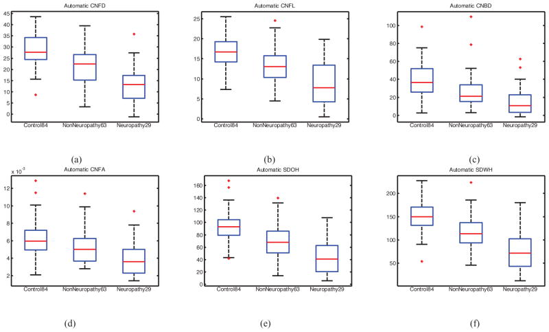
Method: We have evaluated the tool for quantification of Diabetic Sensorimotor Polyneuropathy (DSPN) using both new and previously published morphological features. The evaluation used 888 images from 176 subjects (84 controls and 92 patients with type 1 diabetes). The patient group was further subdivided into those with ( n = 63) and without ( n = 29) DSPN.
Results: We achieve improved nerve- fiber detection over previous results (91.7% sensitivity and specificity in identifying nerve-fiber pixels). Automatic quantification of nerve morphology shows a high correlation with previously reported, manually measured, features. Receiver Operating Characteristic (ROC) analysis of both manual and automatic measurement regimes resulted in similar results in distinguishing patients with DSPN from those without: AUC of about 0.77 and 72% sensitivity-specificity at the equal error rate point.
Conclusion: Automated quantification of corneal nerves in CCM images provides a sensitive tool for identification of DSPN. Its performance is equivalent to manual quantification, while improving speed and repeatability.
Significance: CCM is a novel in vivo imaging modality that has the potential to be a noninvasive and objective image biomarker for peripheral neuropathy. Automatic quantification of nerve morphology is a major step forward in the early diagnosis and assessment of progression, and, in particular, for use in clinical trials to establish therapeutic benefit in diabetic and other peripheral neuropathies.
Figures


References
-
- Boulton AJ. Management of Diabetic Peripheral Neuropathy. Clinical Diabetes. 2005;23(1):9–15.
-
- Daousi C, MacFarlane IA, Woodward A, Nurmikko TJ, Bundred PE, Benbow SJ. Chronic painful peripheral neuropathy in an urban community: a controlled comparison of people with and without diabetes. Diabetic Medicine. 2004;21(9):976–982. - PubMed
-
- Dyck PJ, Overland CJ, Low PA, Litchy WJ, Davies JL, O’Brien PC, Albers JW, Andersen H, Bolton CF, England JD, Klein CJ, Llewelyn JG, Mauermann ML, Russell JW, Singer W, Smith AG, Tesfaye S, Vella A C. v. N. T. Investigators. Signs and symptoms versus nerve conduction studies to diagnose diabetic sensorimotor polyneuropathy: CI vs. NPhys trial. Muscle Nerve. 2010;42(2):157–164. - PMC - PubMed
-
- Dyck PJ, Norell JE, Tritschler H, Schuette K, Samigullin R, Ziegler D, Bastyr EJ, Litchy WJ, O’Brien PC. Challenges in Design of Multicenter Trials: Endpoints Assessed Longitudinally for Change and Monotonicity. Diabetes Care. 2007;30:2619–2625. - PubMed
Publication types
MeSH terms
Grants and funding
LinkOut - more resources
Full Text Sources
Other Literature Sources
Medical
