

Prevalence and Clinicopathological Features of Triaditis in a Prospective Case Series of Symptomatic and Asymptomatic Cats
- PMID: 27296565
- PMCID: PMC5089651
- DOI: 10.1111/jvim.14356
Prevalence and Clinicopathological Features of Triaditis in a Prospective Case Series of Symptomatic and Asymptomatic Cats
Abstract
Background: The term triaditis designates the concurrent presence of idiopathic inflammatory bowel disease (IBD), cholangitis, and pancreatitis in cats.
Hypothesis/objectives: The histopathology of concurrent, but often subclinical, inflammatory processes in the small intestine, liver, and pancreas of cats is poorly described. We aimed to investigate the frequency of enteritis, cholangitis, pancreatitis, or some combination of these in symptomatic and asymptomatic cats, compare clinicopathological features, and correlate histopathological with laboratory findings.
Animals: Domestic cats (27 symptomatic, 20 asymptomatic, and 8 normal).
Methods: Prospective study. Physical examination, laboratory variables (CBC, serum biochemistry profile, serum thyroxine concentration, serum feline trypsin-like immunoreactivity [fTLI], feline lipase immunoreactivity [fPLI, as measured by Spec fPL(®) ], urinalysis, and fecal analysis), imaging, and histopathological examinations were conducted. Feline liver, pancreas, and small intestine were biopsied during laparotomy.
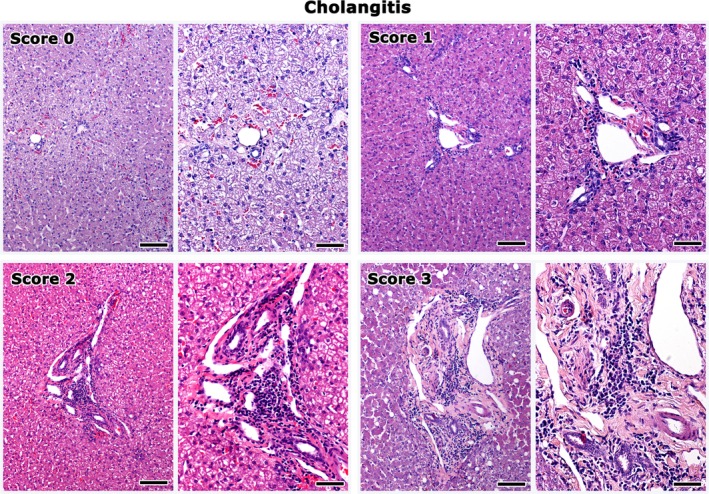
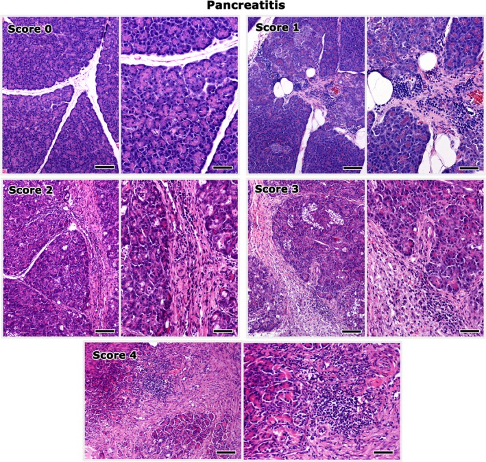
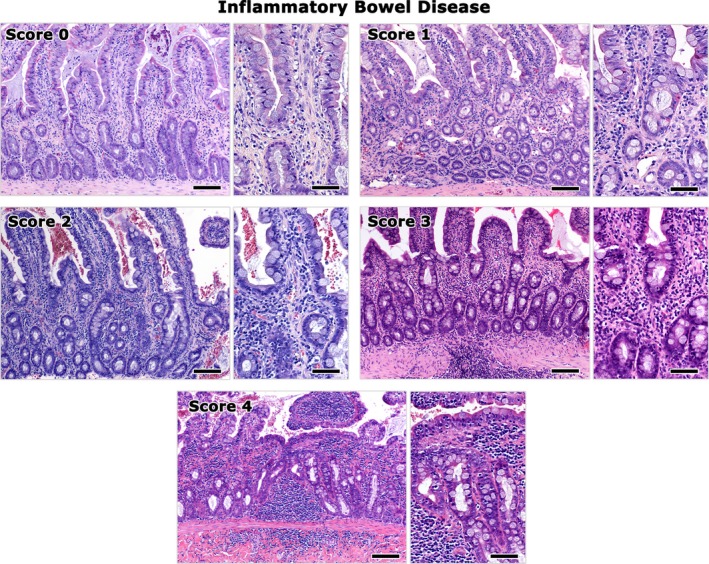
Results: Inflammatory lesions were detected in 47 cats (27 symptomatic, 20 asymptomatic). In total, 20 cats had histopathologic lesions of IBD (13/47, 27.7%), cholangitis (6/47, 12.8%), or pancreatitis (1/47, 2.1%) alone, or inflammation involving >1 organ (27/47, 57.4%). More specifically, 16/47 cats (34.0%) had concurrent lesions of IBD and cholangitis, 3/47 (6.4%) of IBD and pancreatitis, and 8/47 cats (17%) of triaditis. Triaditis was identified only in symptomatic cats (8/27, 29.6%). A mild, positive correlation was detected between the severity (score) of IBD lesions and the number of comorbidities (rho = +0.367, P = .022).
Conclusions and clinical importance: Histopathological evidence of IBD or IBD with comorbidities was detected in both symptomatic and asymptomatic cats. The possibility of triaditis should be considered in symptomatic cats with severe IBD.
Keywords: Cat; Cholangitis; Inflammatory Bowel Disease; Pancreatitis.
Copyright © 2016 The Authors. Journal of Veterinary Internal Medicine published by Wiley Periodicals, Inc. on behalf of the American College of Veterinary Internal Medicine.
Figures
References
-
- Simpson KW. Pancreatitis and triaditis in cats: causes and treatment. J Small Anim Pract 2015;56:40–49. - PubMed
-
- Rothuizen J, Bunch SE, Charles JE, et al., eds. WSAVA Standards for Clinical and Histological Diagnosis of Canine and Feline Liver Diseases: WSAVA Liver Standardization Group. Philadelphia, PA: Elsevier; 2006.
-
- Day MJ, Bilzert T, Mansell J, et al. Histopathological standards for the diagnosis of gastrointestinal inflammation in endoscopic biopsy samples from the dog and cat: A report from the World Small Animal Veterinary Association Gastrointestinal Standardization Group. J Comp Pathol 2008;138:1–40. - PubMed
-
- Washabau RJ, Day MJ, Willard MD, et al. Endoscopic, biopsy, and histopathologic guidelines for the evaluation of gastrointestinal inflammation in companion animals. J Vet Intern Med 2010;24:10–26. - PubMed
MeSH terms
LinkOut - more resources
Full Text Sources
Other Literature Sources
Medical
Miscellaneous