. 2016 Jun 13;4(1):58.
doi: 10.1186/s40478-016-0315-6.

An autoradiographic evaluation of AV-1451 Tau PET in dementia

Affiliations

An autoradiographic evaluation of AV-1451 Tau PET in dementia

Val J Lowe et al. Acta Neuropathol Commun. .

Abstract

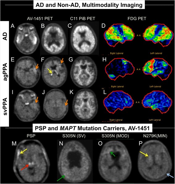
Background: It is essential to determine the specificity of AV-1451 PET for tau in brain imaging by using pathological comparisons. We performed autoradiography in autopsy-confirmed Alzheimer disease and other neurodegenerative disorders to evaluate the specificity of AV-1451 binding for tau aggregates.

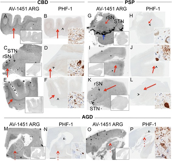
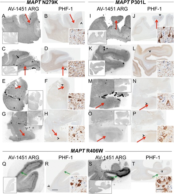
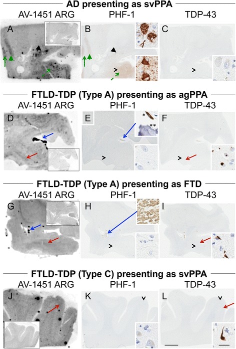
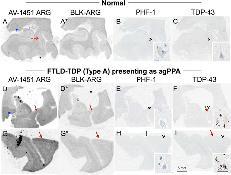
Methods: Tissue samples were selected that had a variety of dementia-related neuropathologies including Alzheimer disease, primary age-related tauopathy, tangle predominant dementia, non-Alzheimer disease tauopathies, frontotemporal dementia, parkinsonism, Lewy body disease and multiple system atrophy (n = 38). Brain tissue sections were stained for tau, TAR DNA-binding protein-43, and α-synuclein and compared to AV-1451 autoradiography on adjacent sections.

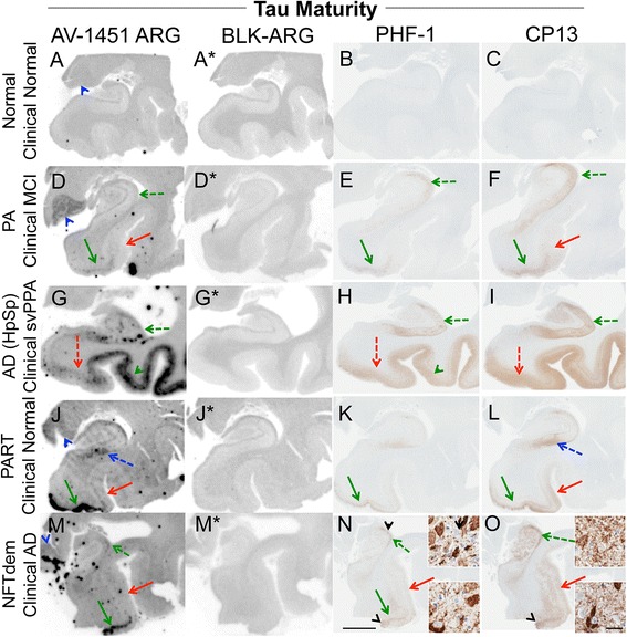
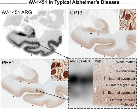
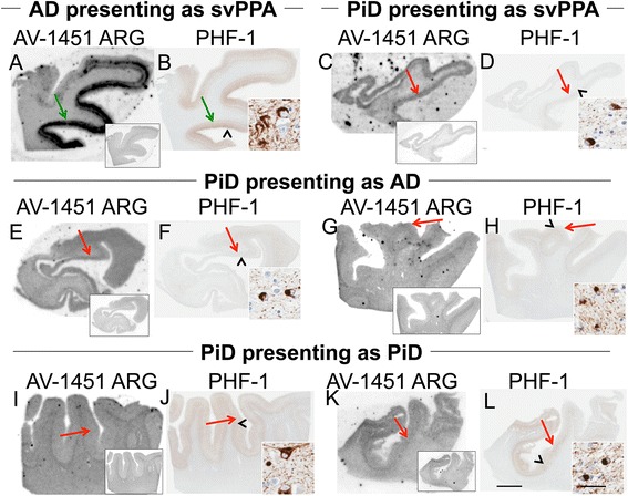
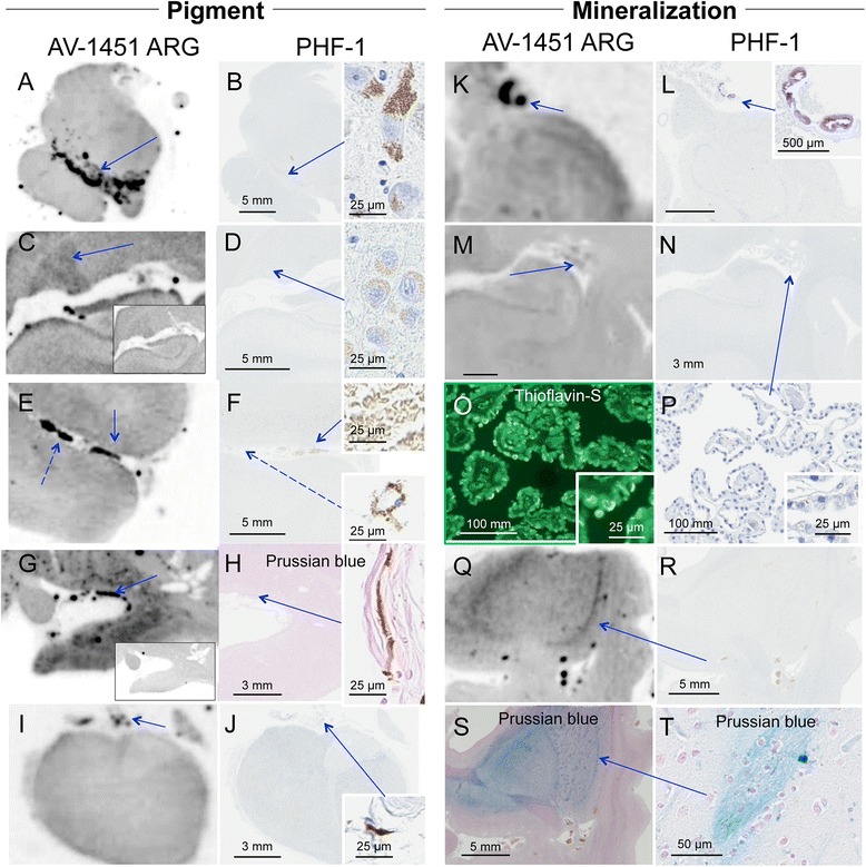
Results: AV-1451 preferentially localized to neurofibrillary tangles, with less binding to areas enriched in neuritic pathology and less mature tau. The strength of AV-1451 binding with respect to tau isoforms in various neurodegenerative disorders was: 3R + 4R tau (e.g., AD) > 3R tau (e.g., Pick disease) or 4R tau. Only minimal binding of AV-1451 to TAR DNA-binding protein-43 positive regions was detected. No binding of AV-1451 to α-synuclein was detected. "Off-target" binding was seen in vessels, iron-associated regions, substantia nigra, calcifications in the choroid plexus, and leptomeningeal melanin.

Conclusions: Reduced AV-1451 binding in neuritic pathology compared to neurofibrillary tangles suggests that the maturity of tau pathology may affect AV-1451 binding and suggests complexity in AV-1451 binding. Poor association of AV-1451 with tauopathies that have preferential accumulation of either 4R tau or 3R tau suggests limited clinical utility in detecting these pathologies. In contrast, for disorders associated with 3R + 4R tau, such as Alzheimer disease, AV-1451 binds tau avidly but does not completely reflect the early stage tau progression suggested by Braak neurofibrillary tangle staging. AV-1451 binding to TAR DNA-binding protein-43 or TAR DNA-binding protein-43 positive regions can be weakly positive. Clinical use of AV-1451 will require a familiarity with distinct types of "off-target" binding.

Keywords: AV-1451; Alzheimer’s disease; Atypical Alzheimer’s disease; Corticobasal degeneration; Frontotemporal dementia; Pick Disease; Pick’s disease; Progressive supranuclear palsy; TDP-43; Tau; Tauopathy.


Figures

Fig. 1
Fig. 1
Examples of AV-1451 PET scans in subjects with various diseases. Correlative PET imaging (AD and Non-AD, Multimodality Imaging, rows 1-3) in an AD subject (1) with high AV-1451 signal (white regions), high PiB signal (white regions) and reduced FDG signal in an “AD” pattern (temporal, parietal and some frontal; colors indicate hypometabolism); an agPPA subject (2) with mild signal in the left temporal and frontal lobes on AV-1451 PET (orange arrows), normal PiB signal, and an “agPPA pattern” on FDG showing left sided dominant frontal and temporal hypometabolism and a svPPA subject (3) with very mild signal in the left temporal lobe on AV-1451 PET (orange arrows), normal PiB signal, and an svPPA pattern on FDG showing left sided dominant temporal hypometabolism. On row 4 (PSP and MAPT Mutation Carriers, AV-1451), from left to right, are AV-1451 PET images showing scattered, minimal signal in tauopathies secondary to PSP and MAPT mutations. The PSP subject showed signal in the brainstem that is likely nonspecific (red arrow). The left MAPT S305N mutation subject has severe (SV) symptoms and secondary ventricular enlargement and the next to the right has moderate (MOD) symptoms. The MAPT N279K mutation subject on the far right has relatively minimal (MIN) symptoms. Some very mildly increased AV-1451 signal is identified in the MAPT S305N subjects diffusely (green arrows) but it is clearly less intense than in the AD subject (1) but only mildly more than in the MAPT N279K subject (blue arrow). Yellow arrows show meningeal (PSP subject), basal ganglia (MAPT N279K subject), and choroid plexus signal (agPPA subject) and are likely non-specific binding regions
Fig. 2
Fig. 2
Correlative AV-1451 autoradiography (ARG) and immunohistochemical (IHC) findings assessing tau maturity, atypical AD and AD tauopathies. (A-C) Case 2 - Normal, (D-F) Case 5 – Pathological aging, (G-I) Case 6 – Hippocampal sparing (HpSp) Alzheimer’s disease (AD), (J-L) Case 10 – Primary Age-Related Tauopathy (PART), (M-O) Case 12 – Neurofibrillary tangle (NFT) predominant dementia. Columns from left to right show AV-1451 ARG, AV-1451 Blocked ARG (BLK-ARG, shown with “letter”*), PHF-1 and CP13 immunohistochemistry in the posterior hippocampal region in cases with various Braak neurofibrillary tangle stages (0-VI) and Thal amyloid phase (0-5). AV-1451 displaceable correspondence with IHC in the medial parahippocampal gyrus (green arrows) is strong with a trend for poorer AV-1451 signal correlation in the lateral parahippocampal gyrus (red arrows) seen especially relative to CP13 except in the hippocampal sparring AD case (G-I). The greatest AV-1451 and IHC concordance was observed in the occiptiotemporal gyrus in AD HpSp (case 6, green arrowhead). There is minimal-moderate AV-1451 signal in the hippocampal formation (CA1, green dashed arrows) that is modest relative to IHC in most samples, again especially as compared to CP13. In the Braak VI AD HpSp case (G-I, case 6), there is minimal AV-1451 signal in the medial parahippocampal gyrus (red dashed arrows) and minimal binding in the hippocampus (green dashed arrows) that is a mismatch to the PHF-1 and CP13. More muted AV-1451 signal was seen in the subiculum in PART (J-K, case 10, blue dashed arrows). Lateral geniculate AV-1451 diffuse, displaceable, minimal signal was seen in many cases (blue arrowheads) and some focal intense signal in some cases (case 10) that may represent vascular non-specific AV-1451 signal. Scale bar for all full size hippocampus at 5 mm, and for 20x zoomed insets at 25 μm. Black arrowheads indicate 20x inset locations
Fig. 3
Fig. 3
Correlative autoradiography (ARG) and immunohistochemical (IHC) findings for assessment of correlative binding in typical Alzheimer’s disease (AD, case 7). AV-1451 ARG (AV-1451 blocking shown in the left ARG inset), PHF-1 and CP13 immunohistochemistry in the posterior hippocampal region in a Braak VI, Thal 5 AD case. Tau pathology in the subiculum is shown in the upper right inset (black arrowhead) and shows very good signal on AV-1451 ARG, PHF-1 and CP13. However, the pattern of AV-1451 binding is similar but not identical to IHC with some focal discordance in many regions including Sommer’s sector of the hippocampus where CA1 and subiculum meet, the superficial layers of the parahippocampal and occipitotemporal gyri (*). Intense, linear AV-1451 signal in cortex matches best with the internal pyramidal cell layer (5) (right lower inset) while superficial layers (1-4) have more minimal-moderate AV-1451 signal where there is intense IHC positivity (right lower inset and yellow arrow). Scale bar for all full size hippocampus at 5 mm, and for 20x zoomed insets at 25 μm. Black arrowheads indicate 20x inset locations
Fig. 4
Fig. 4
Correlative autoradiography (ARG) and immunohistochemical (IHC) findings for assessment of Pick’s disease (PiD, 3R Tau) binding. a, b Case 6 - AD for comparison (HpSp)/DLBD, c, d Case 14 - PiD, e-h Case 15 - PiD, i-l Case 16 – PiD. Shown from left to right AV-1451 ARG (AV-1451 blocking shown in insets) and PHF-1 in the a-f temporal cortex, g-j frontal cortex, and k-l parietal cortex for comparison. Moderate-severe AV-1451 displaceable binding in the AD case is shown (green arrows) corresponding with PHF-1. AV-1451 ARG displaceable signal is minimal and much weaker in the Pick’s cases than in the AD subject even in those with similar intensity of PHF-1 immunopositivity (i-l) to AD (red arrows). In some cases, loss of white matter-grey matter AV-1451 contrast may be the most notable feature of AV-1451 binding as compared to AV-1451 blocking studies (g, i). Scale bar for all full size cortical sections at 5 mm, and for 20x zoomed insets at 25 μm. Black arrowheads indicate 20x inset locations
Fig. 5
Fig. 5
Correlative autoradiography (ARG) and immunohistochemical (IHC) findings for assessment of 4R Tau (CBD, PSP, and AGD) binding. a-d Case 17 – CBD (rostral substantia nigra (rSN), substantia nigra (STN)), e, f Case 18 - CBD, g, h Case 19 - PSP, i-l Case 20 - PSP, m, n Case 22 - AGD, o, p Case 23 – AGD. Shown from left to right AV-1451 ARG (AV-1451 blocking shown in the ARG insets), and PHF-1 IHC in the a, b, e, f frontal and c, d subthalamic regions in CBD cases; g, h midbrain in PSP cases; i, j frontal, k, l subthalamic; and m-p hippocampal regions in AGD cases. AV-1451 displaceable ARG binding corresponding to PHF-1 is often minimal and when present corresponds (red arrows) with the most dense areas of PHF-1 immunopositivity but is overall weaker than in AD subjects. Strong, non-tau related, ARG displaceable signal in the substantia nigra (blue arrow) may be cross-reacting with neuromelanin containing nigral neurons. Minimal displaceable ARG signal in rSN corresponds to PHF-1 immunopositivity (g, k, red arrows). No AV-1451 signal is seen that corresponds to PHF-1 positive AGD (red dashed arrows, m-p). Scale bar for all full size brain sections at 5 mm, and for 20x zoomed insets at 25 μm. Arrowheads indicate 20x inset locations
Fig. 6
Fig. 6
Correlative autoradiography (ARG) and immunohistochemical (IHC) findings for assessment of MAPT mutation (N297K and P3012L) tau binding. a-f Case 24 - FTDP-17 (MAPT N279K), g, h Case 25 - FTDP-17 (MAPT N279K), k-n Case 26 - FTDP-17 (MAPT P301L), i, j, o, p Case 27 - FTDP-17 (MAPT P301L), q-t Case 28 - FTDP-17 (MAPT R406W). Shown from left to right AV-1451 ARG (AV-1451 blocking shown in the ARG insets) and PHF-1 IHC in the a, b, i, j frontal, c, d, k, l, q, r temporal, e, f midbrain, g, h, m-p amygdala, and s, t hippocampus. AV-1451 displaceable ARG signal is minimal but co-localizes in most cases with the most dense areas of PHF-1 immunopositivity (red arrows). Conversely, the MAPT R406W case shows moderate-strong binding in cortical structures with a laminar pattern similar to that observed in AD (green arrows). Scale bar for all full size brain sections at 5 mm, and for 20x zoomed insets at 25 μm. Arrowheads indicate 20x inset locations
Fig. 7
Fig. 7
Correlative autoradiography (ARG) and immunohistochemical (IHC) findings for assessment of TDP-43 binding in cases with different phenotypic TDP-43-like presentations. a-c Case 29 – AD pathologically, d-f Case 30 - FTLD-TDP, g-i Case 31 - FTLD-TDP, j-l Case 32 - FTLD-TDP. Shown from left to right AV-1451 ARG (AV-1451 blocking shown in the ARG insets), and PHF-1 in the a-i amygdala and j-l frontal cortex. a-c A TDP-43-negative AD case presenting clinically as semantic variant of primary progressive aphasia (svPPA) shows strong ARG displaceable binding in insula (green arrow), claustrum (green arrowhead), and NBM (black arrowhead with inset, tangles) and amygdala (black arrow with IHC inset, tangles) with correspondence on PHF-1 IHC. d-f The amygdala region in a FTLD-TDP case presenting as an agrammatic variant of PPA (agPPA) with TDP-43 type A at autopsy, shows a negative PHF-1 and minimal diffuse ARG binding with a weak correspondence (red arrows) with TDP-43 pathology (TDP-43 inset, cytoplasmic and intranuclear inclusions). Strong off-target binding of the vasculature (blue arrow) appears to correspond with mineralization of the vessel and potentially with subpial melanin lentiform-shaped inclusion (upper inset). g, i Similarly, the amygdala of the FTLD-TDP case presenting as FTD with TDP-43 type A at autopsy was negative on PHF-1 and minimally positive on TDP-43. The ARG displaceable binding shows a weak correspondence (red arrows) with TDP-43 pathology (inset). Strong off-target binding of the vasculature (blue arrow) appears to correspond with cross-reactivity to red blood cells (H upper inset). j-l Frontal cortex in a FTLD-TDP case presenting clinically as semantic variant PPA (svPPA) with TDP-43 Type-C at autopsy was negative on PHF-1 and positive on TDP-43. ARG shows minimal displaceable binding in cortex that corresponds (red arrows) to TDP-43 pathology (inset, long thick dystrophic neurite). Scale bar for all full size brain sections at 5 mm, and for 20x zoomed insets at 25 μm. Black arrowheads indicate 20x inset locations
Fig. 8
Fig. 8
Correlative autoradiography (ARG) and immunohistochemical (IHC) findings for assessment of TDP-43 binding. (a-c) Case 2 - Normal, (d-i) Case 30 - FTLD-TDP. Shown from left to right AV-1451 ARG, AV-1451 Blocked ARG (BLK-ARG, shown with “letter”*), PHF-1 and TD-43 in the (a-f) posterior hippocampal and (g-i) superior temporal cortex. ARG images show no AV-1451 in the normal case (a-c) and minimal displaceable AV-1451 uptake in the (d) parahippocampal grey matter and (g) temporal lobe grey matter (red arrows) similar to regions of TDP-43 immunopositivity that is not seen in the normal brain (red dashed arrow, a). Minimal binding of AV-1451 is seen in the lateral geniculate nucleus (blue arrowheads) in both normal and TDP-43 positive cases is likely nonspecific. As an internal control, note that the grey matter parahippocampal AV-1451 signal in the normal case is absent (red dashed arrow) and the lateral geniculate (a) has greater signal—unlike the TDP case (d). Black arrowheads indicate 20x inset locations
Fig. 9
Fig. 9
Correlative autoradiography (ARG) and immunohistochemical (IHC) examples of off-target AV-1451 binding. a, b Case 1 - Normal, c, d Case 3 - Normal, e, f Case 19 - PSP, g, h Case 6 - AD (HpSp), i, j Case 36 - MSA, k, l Case 30 - FTLD-TDP, m-p Case 2 - Normal, q-t Case 31 - FTLD-TDP. We present evidence for two major causes of off-target binding (blue arrows), a-j pigment-laden structures and k-t mineralization. Shown from left to right AV-1451 ARG and PHF-1 IHC (except where noted) are shown in the a, b midbrain, c, d, k-p posterior hippocampus, e, f superior temporal, g, h, q-t amygdala, and i, j pituitary. a, b AV-1451 signal is seen in the substantia nigra in all cases in the study (blue arrow); c, d lateral geniculate displaceable (c, inset) in most cases likely owing to the abundance of pigment-containing lipofuscin-filled neurons (inset); e, f vascular structures on some cases in vessels with cross-reactive red blood cells (top inset); g, h subpial coverings in some cases with strong displaceable (g, inset) binding observed in proximity to melanin-containing structures that were also found on non-antibody based stains (h, Prussian blue, inset); i, j moderate AV-1451 binding in the pituitary section is also found to correspond to subpial melanin. k, l Strong, focal AV-1451 binding was observed in the choroid plexus and found to correspond to mineralization of the vessel (inset). m, n ARG of choroid plexus revealed variable binding with some cases showing little-to-no binding. PHF-1 staining did not reveal tau-positive structures in choroid. Closer inspection of choroid with (o) thioflavin-S fluorescent microscopy revealed abundant Biondi in older brains, which corresponded to (p) pigment-containing structures on zoomed PHF-1 slides. s, t Variable involvement of basal ganglia was noted, but was not found to correspond with tau pathology. s A digitally-modified Prussian blue (iron stain) revealed a similar pattern underlying moderate-strong ARG binding. Curiously, pencil fibers were often noted to be Prussian blue positive. Scale bar for all full size and zoomed brain sections are noted within each image. Black arrowheads indicate 20x inset locations

References

    1. Goedert M, Spillantini MG, Potier MC, Ulrich J, Crowther RA. Cloning and sequencing of the cDNA encoding an isoform of microtubule-associated protein tau containing four tandem repeats: differential expression of tau protein mRNAs in human brain. EMBO J. 1989;8:393–399. - PMC - PubMed
    1. Bennett DA, Schneider JA, Wilson RS, Bienias JL, Arnold SE. Neurofibrillary tangles mediate the association of amyloid load with clinical Alzheimer disease and level of cognitive function. Arch Neurol. 2004;61:378–384. doi: 10.1001/archneur.61.3.378. - DOI - PubMed
    1. Giannakopoulos P, Herrmann FR, Bussiere T, Bouras C, Kovari E, Perl DP, Morrison JH, Gold G, Hof PR. Tangle and neuron numbers, but not amyloid load, predict cognitive status in Alzheimer’s disease. Neurology. 2003;60:1495–1500. doi: 10.1212/01.WNL.0000063311.58879.01. - DOI - PubMed
    1. Janocko NJ, Brodersen KA, Soto-Ortolaza AI, Ross OA, Liesinger AM, Duara R, Graff-Radford NR, Dickson DW, Murray ME. Neuropathologically defined subtypes of Alzheimer’s disease differ significantly from neurofibrillary tangle-predominant dementia. Acta Neuropathol. 2012;124:681–692. doi: 10.1007/s00401-012-1044-y. - DOI - PMC - PubMed
    1. Jellinger KA, Attems J. Neurofibrillary tangle-predominant dementia: comparison with classical Alzheimer disease. Acta Neuropathol. 2007;113:107–117. doi: 10.1007/s00401-006-0156-7. - DOI - PubMed

Publication types

MeSH terms