. 2016 Jul;105(7):2240-8.
doi: 10.1016/j.xphs.2016.05.015. Epub 2016 Jun 11.

Detergent Isolation Stabilizes and Activates the Shigella Type III Secretion System Translocator Protein IpaC

Affiliations

Detergent Isolation Stabilizes and Activates the Shigella Type III Secretion System Translocator Protein IpaC

Abram R Bernard et al. J Pharm Sci. 2016 Jul.

Abstract

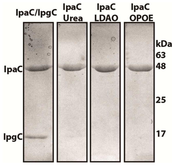
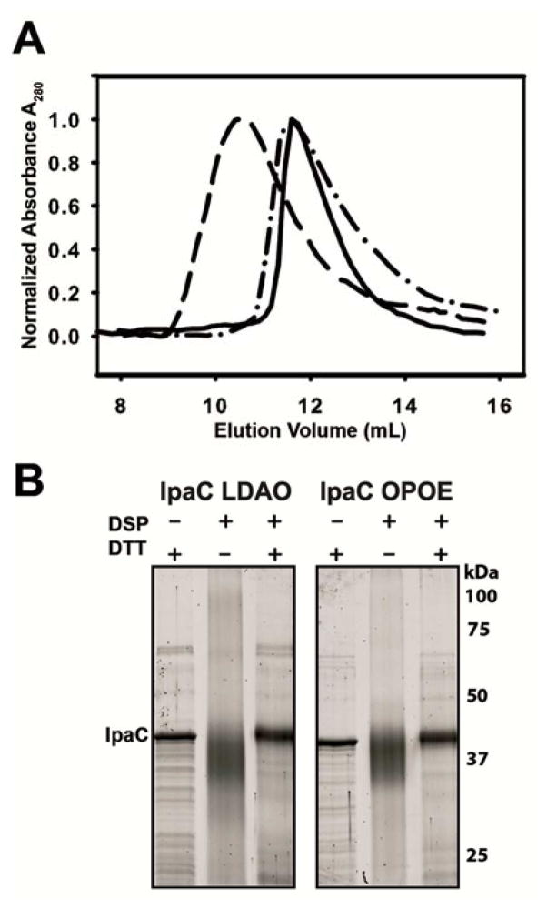
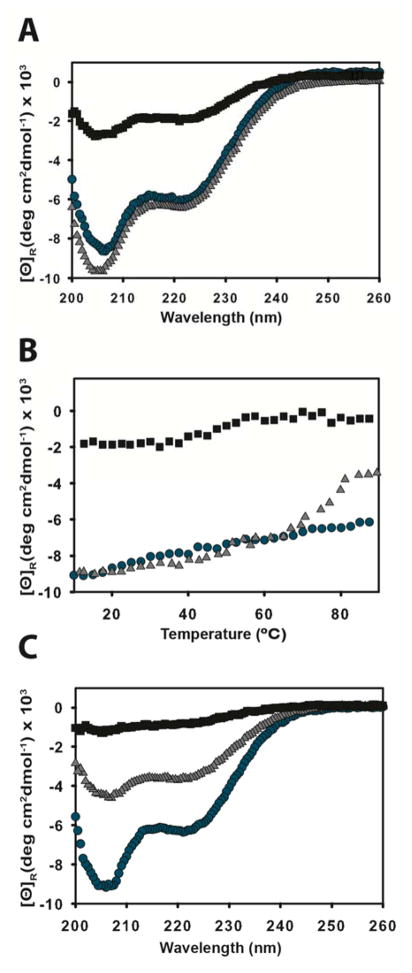
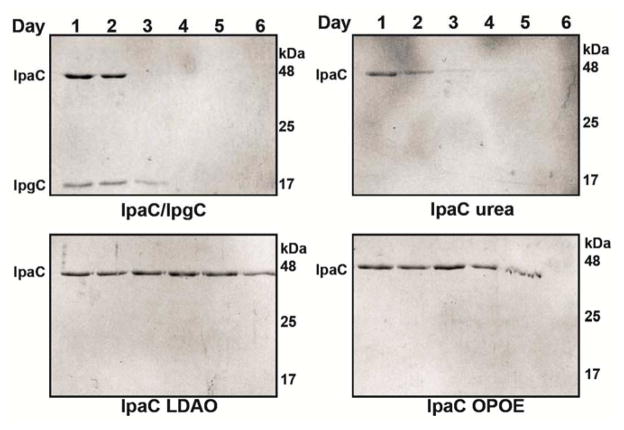
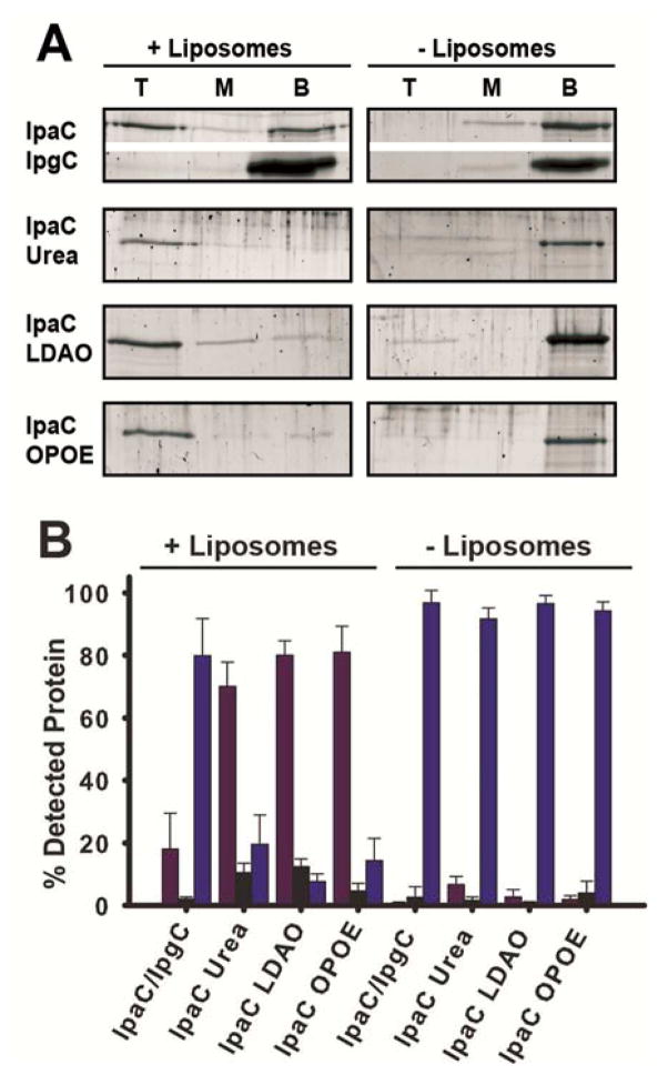
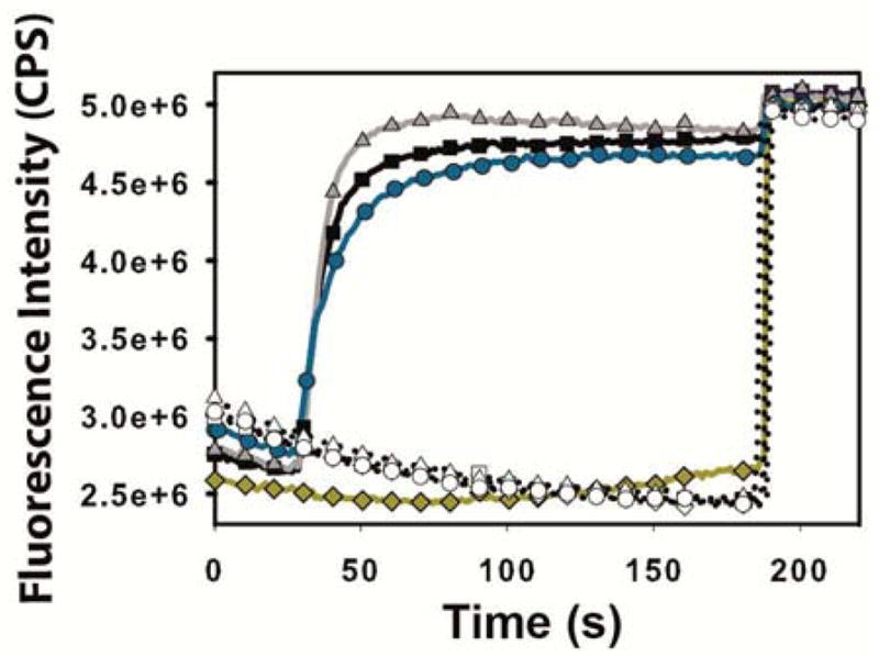
Shigella rely on a type III secretion system as the primary virulence factor for invasion and colonization of human hosts. Although there are an estimated 90 million Shigella infections, annually responsible for more than 100,000 deaths worldwide, challenges isolating and stabilizing many type III secretion system proteins have prevented a full understanding of the Shigella invasion mechanism and additionally slowed progress toward a much needed Shigella vaccine. Here, we show that the non-denaturing zwitterionic detergent N, N-dimethyldodecylamine N-oxide (LDAO) and non-ionic detergent n-octyl-oligo-oxyethylene efficiently isolated the hydrophobic Shigella translocator protein IpaC from the co-purified IpaC/IpgC chaperone-bound complex. Both detergents resulted in monomeric IpaC that exhibits strong membrane binding and lysis characteristics while the chaperone-bound complex does not, suggesting that the stabilizing detergents provide a means of following IpaC "activation" in vitro. Additionally, biophysical characterization found that LDAO provides significant thermal and temporal stability to IpaC, protecting it for several days at room temperature and brief exposure to temperatures reaching 90°C. In summary, this work identified and characterized conditions that provide stable, membrane active IpaC, providing insight into key interactions with membranes and laying a strong foundation for future vaccine formulation studies taking advantage of the native immunogenicity of IpaC and the stability provided by LDAO.

Keywords: circular dichroism; light scattering (dynamic); liposomes; physical characterization; physical stability.


Conflict of interest statement

CONFLICT OF INTEREST

The authors declare no personal financial or non-financial conflicts of interest.

Figures

Figure 1
Figure 1. Detergent isolation of IpaC from stable co-purified IpaC/IpgC complexes
SDS-PAGE gel showing the highly pure IpaC/IpgC complex resulting from co-purification with Ni2+ chelation chromatography targeting the N-terminal 6X histidine tag on IpaC followed by size exclusion chromatography of the complex. The purified IpaC/IpgC was re-bound to a Ni2+ column and IpgC removed by washing the column with binding buffer containing 6 M Urea, 0.1% LDAO or 1.0% OPOE to disrupt the IpaC/IpgC complex prior to elution of IpaC in buffer containing 400 mM imidazole and 2 M Urea, 0.05% LDAO or 0.5% OPOE to stabilize the isolated IpaC (left to right).
Figure 2
Figure 2. IpaC remains monomeric in LDAO and OPOE
A) Size exclusion chromatography analysis of IpaC in 2 M urea (dash), 0.5% OPOE (dot dash) and 0.05% LDAO (solid). Urea stabilized IpaC results in a main elution peak centered at 0.44 column volumes while IpaC in either 0.5% OPOE or 0.05% LDAO exhibit similar profiles that elute at approximately 0.49 column volumes. B) SDS-PAGE analysis of DSP crosslinked IpaC in 0.05% LDAO (left 3 lanes) and in 0.5% OPOE (right 3 lanes). The addition of DSP shows that IpaC is monomeric in both LDAO and OPOE as the protein remains centered at the 42 kDa denatured and reduced species in the first lane. The broadened band in the crosslinked IpaC conditions results from intramolecular crosslinking that is relieved upon the cleavage of the DSP internal disulfide with DTT (right lanes).
Figure 3
Figure 3. CD analysis of IpaC secondary structure content and thermal stability
A) IpaC was analyzed in the presence of 200 mM urea, 0.05% LDAO and 0.5% OPOE (black squares, blue circles and gray triangles respectively) showing that IpaC in both detergents exhibits strong CD profiles while urea results in significant unfolding of the protein. B) Thermal unfolding of IpaC prepared in 2 M urea, 0.05% LDAO and 0.5% OPOE (black squares, blue circles and gray triangles respectively) is shown based on monitoring the CD signal at 208 nm as the sample was heated from 10°C to 90°C. C) Far-UV CD scans of IpaC in 200 mM urea, 0.05% LDAO and 0.5% OPOE (black squares, blue circles and gray triangles respectively) following the thermal melts displayed in panel B and return to 10°C, showing that IpaC in LDAO maintained nearly all native secondary structure content following heating to 90°C.
Figure 4
Figure 4. LDAO and OPOE enhance the temporal stability of recombinant IpaC
The co-purified IpaC/IpgC complex and isolated IpaC in 2 M urea, 0.05% LDAO, or 0.5% OPOE was maintained at room temperature and sampled for 6 days. The daily samples were analyzed by SDS-PAGE for both protein levels and degradation. The IpaC/IpgC complex and IpaC in 2 M urea are no longer observed in solution after 2 days (coincides with observed protein precipitation). Both OPOE and LDAO enhanced IpaC stability with IpaC in LDAO showing little change over the time course of the experiment.
Figure 5
Figure 5. Liposome flotation indicates that IpgC association prevents IpaC interaction with liposomes while IpaC isolation promotes liposome binding
Liposome flotation was carried out as described in the Methods section. Protein interacting with liposomes migrates to the top fraction while protein unable to bind liposomes remains in the bottom fraction of the sucrose gradient. The presence of protein in top, middle, and bottom fractions of the sucrose gradient was determined by SDS–PAGE and quantified by Oriole total protein staining followed by densitometry analysis. A) Representative gels used to determine the location of IpaC within the sucrose gradient. B) Quantitative densitometry results identifying the position of all tested IpaC conditions within the sucrose gradient. Red bars represent the percentage of the total protein in the top fraction of the sucrose gradient, black bars represent the percentage in the middle fraction, and blue is the relative amount of protein that remained in the bottom fraction for each condition. The total amount of protein for each condition totals 100% and is plotted as the mean ± SD of 3 independent analyses. Co-purified IpaC/IpgC did not interact with liposomes and remained predominantly in the bottom gradient fraction while IpaC isolated from IpgC by urea, OPOE, or LDAO all efficiently bound liposomes and migrated to the top fraction of the sucrose gradient. In the absence of liposomes, all conditions resulted in the protein remaining in the bottom fraction.
Figure 6
Figure 6. Isolated recombinant IpaC promotes SRB release from liposomes
Disruption of liposomes and release of encapsulated SRB relieves autoquenching and results in an increase in rhodamine fluorescence emission. Representative time-dependent fluorescence curves following liposome exposure to tested conditions containing IpaC (filled shapes) and corresponding buffer/detergent only controls (hollow shapes). IpaC/IpgC complex in PBS, yellow diamond; IpaC in PBS with 2 M urea (final [urea] ≤ 10 mM), black square; IpaC in PBS with 0.05% LDAO, blue circle; IpaC in PBS with 0.5% OPOE, gray triangle. The protein (or buffer control) was added seconds after fluorescence collection was initiated and Triton X-100 added after 180 seconds to fully disrupt remaining liposomes and provide a 100% lysis benchmark for quantitation of dye release.

References

    1. Schroeder GN, Hilbi H. Molecular pathogenesis of Shigella spp: controlling host cell signaling, invasion, and death by type III secretion. Clinical microbiology reviews. 2008;21:134–156. - PMC - PubMed
    1. WHO. Initiative for Vaccine Research (IVR) Diarrhoeal Diseases. 2009 http://www.who.int/vaccine_research/diseases?diarrhoeal/en/index6.html.
    1. DuPont HL, Levine MM, Hornick RB, Formal SB. Inoculum size in shigellosis and implications for expected mode of transmission. The Journal of infectious diseases. 1989;159:1126–1128. - PubMed
    1. Dutta S, Ghosh A, Ghosh K, Dutta D, Bhattacharya SK, Nair GB, Yoshida S. Newly emerged multiple-antibiotic-resistant Shigella dysenteriae type 1 strains in and around Kolkata, India, are clonal. Journal of clinical microbiology. 2003;41:5833–5834. - PMC - PubMed
    1. Wassef JS, Keren DF, Mailloux JL. Role of M cells in initial antigen uptake and in ulcer formation in the rabbit intestinal loop model of shigellosis. Infection and immunity. 1989;57:858–863. - PMC - PubMed

Publication types