

Echocardiographic Linear Dimensions for Assessment of Right Ventricular Chamber Volume as Demonstrated by Cardiac Magnetic Resonance
- PMID: 27297619
- PMCID: PMC5057385
- DOI: 10.1016/j.echo.2016.05.002
Echocardiographic Linear Dimensions for Assessment of Right Ventricular Chamber Volume as Demonstrated by Cardiac Magnetic Resonance
Abstract
Background: Echocardiography-derived linear dimensions offer straightforward indices of right ventricular (RV) structure but have not been systematically compared with RV volumes on cardiac magnetic resonance (CMR).
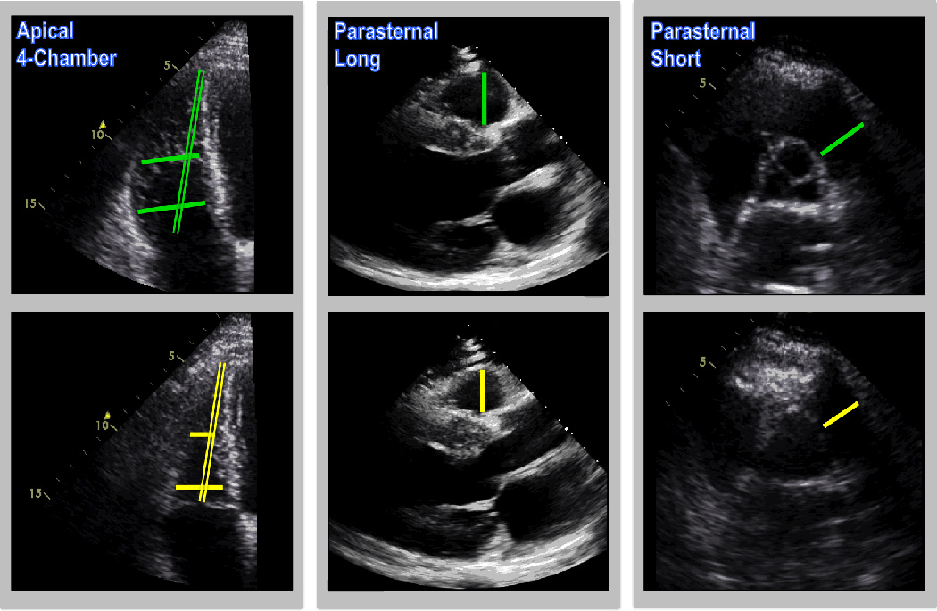
Methods: Echocardiography and CMR were interpreted among patients with coronary artery disease imaged via prospective (90%) and retrospective (10%) registries. For echocardiography, American Society of Echocardiography-recommended RV dimensions were measured in apical four-chamber (basal RV width, mid RV width, and RV length), parasternal long-axis (proximal RV outflow tract [RVOT]), and short-axis (distal RVOT) views. For CMR, RV end-diastolic volume and RV end-systolic volume were quantified using border planimetry.
Results: Two hundred seventy-two patients underwent echocardiography and CMR within a narrow interval (0.4 ± 1.0 days); complete acquisition of all American Society of Echocardiography-recommended dimensions was feasible in 98%. All echocardiographic dimensions differed between patients with and those without RV dilation on CMR (P < .05). Basal RV width (r = 0.70), proximal RVOT width (r = 0.68), and RV length (r = 0.61) yielded the highest correlations with RV end-diastolic volume on CMR; end-systolic dimensions yielded similar correlations (r = 0.68, r = 0.66, and r = 0.65, respectively). In multivariate regression, basal RV width (regression coefficient = 1.96 per mm; 95% CI, 1.22-2.70; P < .001), RV length (regression coefficient = 0.97; 95% CI, 0.56-1.37; P < .001), and proximal RVOT width (regression coefficient = 2.62; 95% CI, 1.79-3.44; P < .001) were independently associated with CMR RV end-diastolic volume (r = 0.80). RV end-systolic volume was similarly associated with echocardiographic dimensions (basal RV width: 1.59 per mm [95% CI, 1.06-2.13], P < .001; RV length: 1.00 [95% CI, 0.66-1.34], P < .001; proximal RVOT width: 1.80 [95% CI, 1.22-2.39], P < .001) (r = 0.79).
Conclusions: RV linear dimensions provide readily obtainable markers of RV chamber size. Proximal RVOT and basal width are independently associated with CMR volumes, supporting the use of multiple linear dimensions when assessing RV size on echocardiography.
Keywords: Cardiovascular magnetic resonance; Echocardiography; Right ventricle.
Copyright © 2016 American Society of Echocardiography. Published by Elsevier Inc. All rights reserved.
Conflict of interest statement
Conflicts of Interests Disclosure: None
Figures

References
-
- Anavekar NS, Skali H, Bourgoun M, Ghali JK, Kober L, Maggioni AP, et al. Usefulness of right ventricular fractional area change to predict death, heart failure, and stroke following myocardial infarction (from the VALIANT ECHO Study) Am J Cardiol. 2008;101:607–612. - PubMed
-
- Antoni ML, Scherptong RW, Atary JZ, Boersma E, Holman ER, van der Wall EE, et al. Prognostic value of right ventricular function in patients after acute myocardial infarction treated with primary percutaneous coronary intervention. Circ Cardiovasc Imaging. 2010;3:264–271. - PubMed
-
- Lang RM, Badano LP, Mor-Avi V, Afilalo J, Armstrong A, Ernande L, et al. Recommendations for cardiac chamber quantification by echocardiography in adults: an update from the American Society of Echocardiography and the European Association of Cardiovascular Imaging. J Am Soc Echocardiogr. 2015;28:1–39. e14. - PubMed
-
- Devereux RB, Reichek N. Echocardiographic determination of left ventricular mass in man. Anatomic validation of the method. Circulation. 1977;55:613–618. - PubMed
-
- Devereux RB, Roman MJ, Palmieri V, Liu JE, Lee ET, Best LG, et al. Prognostic implications of ejection fraction from linear echocardiographic dimensions: the Strong Heart Study. Am Heart J. 2003;146:527–534. - PubMed
Publication types
MeSH terms
Grants and funding
LinkOut - more resources
Full Text Sources
Other Literature Sources
Medical
