. 2016 Jun 14:6:27941.
doi: 10.1038/srep27941.

Next generation bone tissue engineering: non-viral miR-133a inhibition using collagen-nanohydroxyapatite scaffolds rapidly enhances osteogenesis

Affiliations

Next generation bone tissue engineering: non-viral miR-133a inhibition using collagen-nanohydroxyapatite scaffolds rapidly enhances osteogenesis

Irene Mencía Castaño et al. Sci Rep. .

Abstract

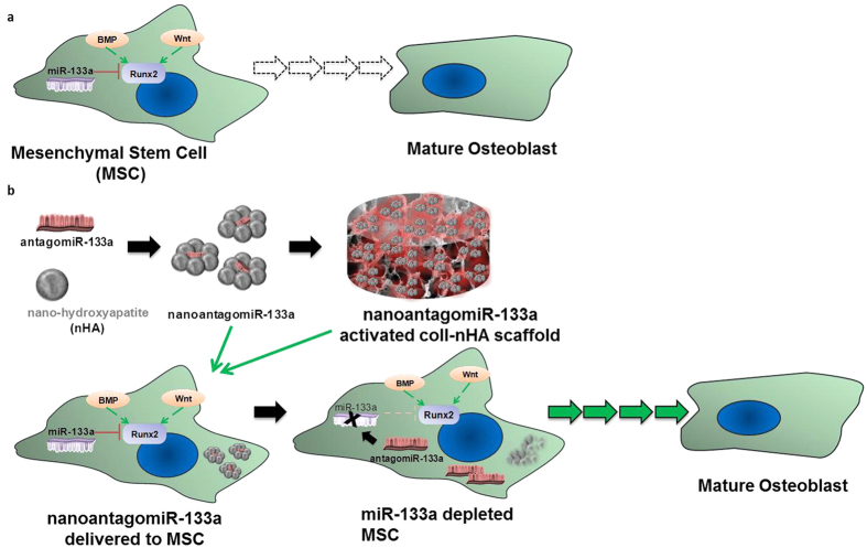
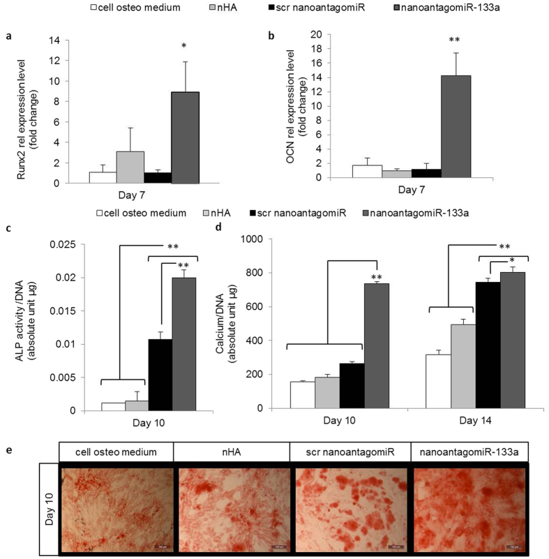
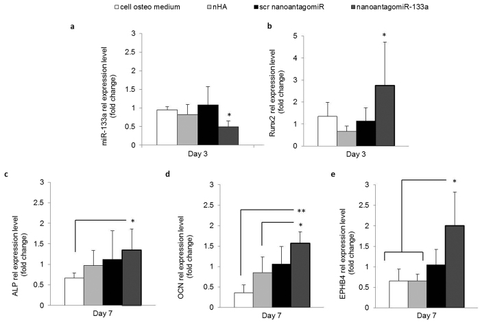
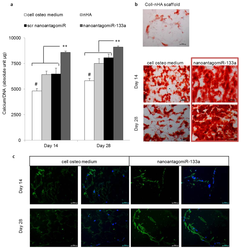
Bone grafts are the second most transplanted materials worldwide at a global cost to healthcare systems valued over $30 billion every year. The influence of microRNAs in the regenerative capacity of stem cells offers vast therapeutic potential towards bone grafting; however their efficient delivery to the target site remains a major challenge. This study describes how the functionalisation of porous collagen-nanohydroxyapatite (nHA) scaffolds with miR-133a inhibiting complexes, delivered using non-viral nHA particles, enhanced human mesenchymal stem cell-mediated osteogenesis through the novel focus on a key activator of osteogenesis, Runx2. This study showed enhanced Runx2 and osteocalcin expression, as well as increased alkaline phosphatase activity and calcium deposition, thus demonstrating a further enhanced therapeutic potential of a biomaterial previously optimised for bone repair applications. The promising features of this platform offer potential for a myriad of applications beyond bone repair and tissue engineering, thus presenting a new paradigm for microRNA-based therapeutics.


Figures

Figure 1
Figure 1. AntagomiR-133a role in mesenchymal stem cell (MSC) osteodifferentiation.
(a) Extracellular ligands such as Bone Morphogenetic Proteins (BMPs) and wingless-related integration site family (Wnt) proteins initiate complex signaling pathways (green arrows) that activate the Runt-related transcription factor 2 (Runx2) to initiate differentiation towards a mature osteoblast state, while miR-133a specifically targets and inhibits (red brake symbol) Runx2. (b) AntagomiR-133a forms complexes with nHA particles which are either delivered to MSCs directly or on porous collagen-nHA scaffolds. They bind to and inhibit miR-133a (black arrow and X symbol), diminishing the silencing of Runx2 (faded red brake symbol), which results in higher availability of functional levels of Runx2. Runx2 drives MSCs along the osteogenic lineage in progressive maturity stages.
Figure 2
Figure 2. qRT-PCR analysis of miR-133a role in hMSC osteogenesis.
Comparison of miR-133a intracellular levels between cells cultured in standard growth medium versus osteogenic media over the course of 14 days demonstrated a natural decrease in miR-133a at later timepoints in osteogenic culture. Mean + standard deviation, n = 4, *p < 0.05, **p < 0.001.
Figure 3
Figure 3. qRT-PCR analysis of miRNA manipulation in hMSC monolayer cultured in osteogenic medium.
(a) NanoantagomiR-133a treatment demonstrated a maintained functionality with high silencing of miR-133a intracellular levels in hMSC monolayer osteogenic culture. (b) NanoantagomiR-16 treatment did not modify intracellular miR-133a levels and (c) nanoantagomiR-133a treatment did not modify intracellular miR-16 levels demonstrating treatment specificity and indicating no RISC overloading associated effects. Mean + standard deviation, n = 4, *p < 0.05, **p < 0.001, NS = not significant variation.
Figure 4
Figure 4. NanoantagomiR-133a treatment enhanced osteogenesis markers in hMSC monolayer.
(a)Runx2 mRNA expression and (b) OCN mRNA relative level were increased in the nanoantagomiR-133a group after 7 days. (c) Significantly increased ALP activity levels were found in the nanoantagomiR-133a group 10 days after treatment. Mean + standard deviation, n = 3, **p < 0.001. (d) Calcium deposition was markedly increased by day 10 and maintained increased calcium levels compared to the control groups at 14 days after treatment with nanoantagomiR-133a in osteogenic culture. (e) Alizarin red staining showed calcium deposits at 10 days after treatment, scale bar = 100 μm. Mean + standard deviation, (a,b) n = 4, (c,d) n = 3, *p < 0.05, **p < 0.001.
Figure 5
Figure 5. Enhanced osteogenic markers in hMSC 3D culture.
(a) miR-133a intracellular levels were significantly decreased for hMSCs cultured on the nanoantagomiR-133a activated scaffolds in comparison to the scr activated scaffolds over a timecourse of 14 days, demonstrating a high silencing functionality of the non-viral based 3D delivery system. (b) Runx2 mRNA expression was upregulated in the nanoantagomiR-133a activated scaffold group after 3 days. Mean + standard deviation, n = 5, *p < 0.05. (c) ALP, (d) OCN and (e) EPHB4 mRNA expression was upregulated in the nanoantagomiR-133a activated scaffold group after 7 days. Mean + standard deviation, n = 4, *p < 0.05, **p < 0.001.
Figure 6
Figure 6. Enhanced mineral matrix deposition in hMSC 3D culture.
(a) Calcium normalised to dsDNA content confirmed a significant increase in calcium deposition by day 14 in nanoantagomiR-133a activated coll-nHA scaffolds and maintained increased calcium levels compared to the control groups after 28 days. Non cell-seeded scaffolds were used as a control for the determination of calcium presence in the extracellular matrix. Mean + standard deviation, n = 3, **p < 0.001, #p < 0.001 compared to all other groups. (b) Alizarin red staining showed increased calcium deposits in the nanoantagomiR-133a loaded coll-nHA scaffold group at 14 and 28 days compared to all other groups. Scale bar = 50 μm. (c) OCN immunofluorescence staining (green) after 14 and 28 days in 3D osteogenic culture showed increased protein expression in the nanoantagomiR-133a loaded scaffolds in comparison with the control treatment groups. Nuclei depicted in blue, for DAPI staining, Scale bar = 50 μm.

References

    1. Elmore J. C., Larsen C. & Neptune C. U. S. Markets for musculoskeletal tissue engineering and cell transplantation products. Report No. A422, Market and technology Reports (Medtech Insights, New York, 2010).
    1. Gleeson J. P., Plunkett N. A. & O’Brien F. J. Addition of hydroxyapatite improves stiffness, interconnectivity and osteogenic potential of a highly porous collagen-based scaffold for bone tissue regeneration. Eur. Cells Mater. 20, 218–230 (2010). - PubMed
    1. Clark E. A., Kalomoiris S., Nolta J. A. & Fierro F. A. Concise review: Microrna function in multipotent mesenchymal stromal cells. Stem Cells 32, 1074–1082 (2014). - PMC - PubMed
    1. Beavers K. R., Nelson C. E. & Duvall C. L. Mirna inhibition in tissue engineering and regenerative medicine. Adv. Drug Delivery Rev. 88, 123–137 (2015). - PMC - PubMed
    1. Hu R., Li H., Liu W., Yang L., Tan Y. & Luo X. Targeting mirnas in osteoblast differentiation and bone formation. Expert Opin. Ther. Targets 14, 1109–1120 (2010). - PubMed

Publication types

MeSH terms