. 2016 Aug 1;416(1):111-122.
doi: 10.1016/j.ydbio.2016.05.038. Epub 2016 Jun 10.

Genetic and biochemical evidence that gastrulation defects in Pofut2 mutants result from defects in ADAMTS9 secretion

Affiliations

Genetic and biochemical evidence that gastrulation defects in Pofut2 mutants result from defects in ADAMTS9 secretion

Brian A Benz et al. Dev Biol. .

Abstract

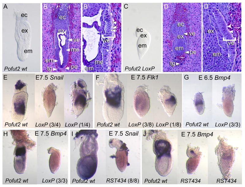
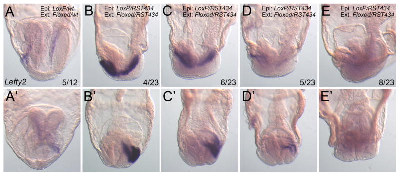
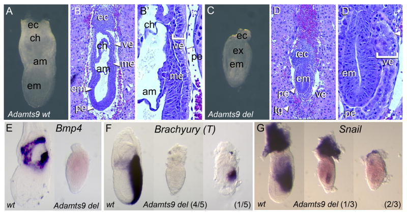
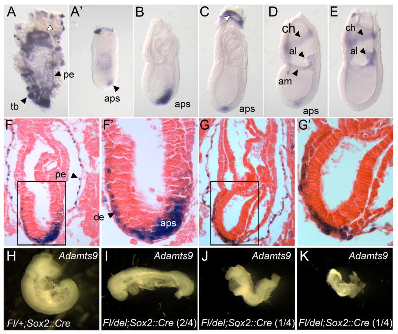
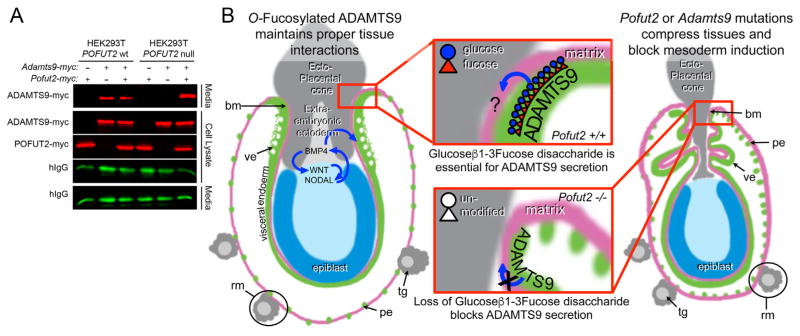
Protein O-fucosyltransferase 2 (POFUT2) adds O-linked fucose to Thrombospondin Type 1 Repeats (TSR) in 49 potential target proteins. Nearly half the POFUT2 targets belong to the A Disintegrin and Metalloprotease with ThromboSpondin type-1 motifs (ADAMTS) or ADAMTS-like family of proteins. Both the mouse Pofut2 RST434 gene trap allele and the Adamts9 knockout were reported to result in early embryonic lethality, suggesting that defects in Pofut2 mutant embryos could result from loss of O-fucosylation on ADAMTS9. To address this question, we compared the Pofut2 and Adamts9 knockout phenotypes and used Cre-mediated deletion of Pofut2 and Adamts9 to dissect the tissue-specific role of O-fucosylated ADAMTS9 during gastrulation. Disruption of Pofut2 using the knockout (LoxP) or gene trap (RST434) allele, as well as deletion of Adamts9, resulted in disorganized epithelia (epiblast, extraembryonic ectoderm, and visceral endoderm) and blocked mesoderm formation during gastrulation. The similarity between Pofut2 and Adamts9 mutants suggested that disruption of ADAMTS9 function could be responsible for the gastrulation defects observed in Pofut2 mutants. Consistent with this prediction, CRISPR/Cas9 knockout of POFUT2 in HEK293T cells blocked secretion of ADAMTS9. We determined that Adamts9 was dynamically expressed during mouse gastrulation by trophoblast giant cells, parietal endoderm, the most proximal visceral endoderm adjacent to the ectoplacental cone, extraembryonic mesoderm, and anterior primitive streak. Conditional deletion of either Pofut2 or Adamts9 in the epiblast rescues the gastrulation defects, and identified a new role for O-fucosylated ADAMTS9 during morphogenesis of the amnion and axial mesendoderm. Combined, these results suggested that loss of ADAMTS9 function in the extra embryonic tissue is responsible for gastrulation defects in the Pofut2 knockout. We hypothesize that loss of ADAMTS9 function in the most proximal visceral endoderm leads to slippage of the visceral endoderm and altered characteristics of the extraembryonic ectoderm. Consequently, loss of input from the extraembryonic ectoderm and/or compression of the epiblast by Reichert's membrane blocks gastrulation. In the future, the Pofut2 and Adamts9 knockouts will be valuable tools for understanding how local changes in the properties of the extracellular matrix influence the organization of tissues during mammalian development.

Keywords: Adamts9; Gastrulation; O-Fucosylation; Pofut2; Thrombospondin type I repeats.


Figures

Fig. 1
Fig. 1
POFUT2 is essential for maintaining epithelial organization and mesoderm differentiation. (A–D′) Comparison of E 7.5 Pofut2 wild-type (Pofut2 wt) and Pofut2 knockout embryos (Pofut2-LoxP) by gross morphology (A and C) and hematoxylin and eosin staining of sections (B and D). Embryo orientation: proximal up and distal down; anterior left and posterior right. (A–B′) wild-type embryos have well-defined embryonic (embryonic ectoderm, ec; mesoderm, me) and extraembryonic tissues (ectoplacental cone, ec; extraembryonic ectoderm, ex; embryonic ectoderm, em; visceral endoderm, ve; parietal endoderm, pe; trophoblast giant cells, tg). (B′) The proximal visceral endoderm (bracketed) is highly polarized and has large apical vacuoles (arrowheads). (C–D′) In contrast, tissues in Pofut2 knockout embryos are highly compressed, mesoderm is not clearly visible, and extraembryonic structures derived from mesoderm are absent. (D′) The Pofut2 knockout visceral endoderm, although polarized, lacks large apical vacuoles (arrowheads). (E–J) Comparison of mesodermal (Snail and Flk1) and mesodermal and extraembryonic ectoderm (Bmp4) marker expression using whole mount in situ hybridization. Where the expression patterns varied in Pofut2 mutants, representative embryos are shown with number of embryos in indicated in parenthesis. (E–F) Comparison of gene expression in wild-type (left) and Pofut2-LoxP mutants (right). In contrast to E 7.5 wild-type embryos, the majority of Pofut2 mutants fail to express these genes, or expressed them at lower levels. Note that 4/8 Pofut2-LoxP mutants did not express Flk1 (not shown). (I, J) Comparison of Snail (I) and Bmp4 (J) expression in wild-type (left) and Pofut2-RST434 mutant littermates (right). Pofut2-RST434 mutants lack Snail mRNA and show only limited expression of Bmp4 in the extraembryonic region. In this study, the Pofut2-RST434 had been back-crossed more than 20 generations to C57BL/6J. Embryos are oriented proximal up and distal down. Wild-type embryos are oriented anterior to the left; Anterior posterior orientation of Pofut2 mutants is not known.
Fig. 2
Fig. 2
Pofut2 activity in the epiblast is essential for axis elongation. The genotypes of the epiblast (Epi) and extraembryonic (Ext) tissues in the embryo pairs (control on the left and epiblast mutants on the right) are indicated at the top of the figure. To evaluate the effects of deleting Pofut2 in the epiblast using Sox2::Cre, embryos were obtained between E 7.5 (A–B, G) and 8.5 (C–F, H) and were processed for whole-mount in situ hybridization expression or were embedded, sectioned, and stained with hematoxylin and eosin to evaluate tissue morphology (G and H). (A, C, and E) T was expressed in the primitive streak and notochord of Pofut2 wild-type and epiblast mutants, suggesting that loss of POFUT2 activity in the extraembryonic tissue was responsible for blocking gastrulation in the Pofut2 mutants (Fig. 1). Arrowheads in panel E identify discontinuous notochord. (E) Inset shows PCR genotyping of embryos. Lanes correspond to embryo on the left and right. Full litters for panels C and E are shown in Supplementary Fig. 3 with complete genotyping. (B, D, and F) Foxa2 was expressed in the anterior primitive streak and emerging mesendoderm at the late primitive streak stage (B), notochord at the early headfold stage (D), and notochord and floorplate at late headfold stage (F) in Pofut2 wild-type and epiblast mutants. Both T and Foxa2 expression domains were wider and somewhat irregular in shape in the Pofut2 epiblast mutants compared to wild-type littermates. (G and H) Sagittal sections of Pofut2 wild-type and epiblast mutant littermates at E 7.5 (G) E 8.5 (H) stained with hematoxylin and eosin. (G) At E 7.5 (late bud stage), the chorion frequently remains attached to the amnion in Pofut2 epiblast mutants and the epiblast appears disorganized. Persistent attachment of chorion and amnion is indicated by asterisk (*). (H) At E 8.5, there is evidence of heart and somite development in Pofut2 epiblast mutants. Anterior is to the left, posterior to the right, proximal up, and distal down. Amnion, am; allantois, al; blood islands, bl; brachial arch, ba; brain, br; chorion, ch; heart, he; headfold, hf; neural fold, nf; node, n; notochord, nt; neural tube, nr; optic vesicle, ov; somite, so.
Fig. 3
Fig. 3
Laterality is likely established in Pofut2 epiblast mutants. Representative whole mount Lefty2 in situ hybridization in E8.5 embryos viewed from the left side (anterior to the left) (A–E) or the anterior (A′–E′). The number of Pofut2 wild-type (Pofut2 wt) or Pofut2 epiblast mutant (Pofut2 epi mut) embryos displaying the respective expression pattern is shown as a fraction of total embryos analyzed. (A) In wild-type embryos Lefty2 was expressed in the lateral plate mesoderm or 5 embryos and extinguished in 7. (B–E) A range of Lefty2 expression in the lateral plate mesoderm was detected in the majority of Pofut2 epiblast mutant embryos. Genotype of epiblast (Epi:) and extraembryonic tissues (Ext:) are shown for each embryo.
Fig. 4
Fig. 4
Adamts9 null embryos recapitulate the Pofut2 knockout phenotype. (A–D′) Comparison of gross morphology of E 7.5 Adamts9 wild-type (Adamts9 wt) and Adamts9 nullembryos (Adamts9del/del) (A and C) and after hematoxylin and eosin staining of sections (B and D). Wild-type embryos (A and B) are similar to those described in Fig. 1(A–B′). In contrast, the epithelia of Adamts9 mutants (C–D) were disorganized and the visceral endoderm lacked apical vacuoles, similar to that observed in Pofut2 knockouts (Fig. 1C–D′). White arrowheads in panel D′ point to the absence of apical vesicles in Adamts9 mutants. (E–G) Whole-mount in situ hybridization comparison of Adamts9 wild-type and knockout embryos at E 7.5. (E) Bmp4 was not expressed in Adamts9 mutants, similar to that observed in the Pofut2 knockout (Fig. 1G). (F) Brachyury (T) was expressed in a truncated domain in 1/5 Adamts9 mutants, but was absent in 4/5 embryos. (G) Snail was expressed weakly in 1/3 Adamts9 mutants. Embryos are oriented proximal up and distal down. Wild-type embryos are oriented anterior to the left and posterior to the right. The anterior posterior orientation of Pofut2 mutants is not known.
Fig. 5
Fig. 5
Adamts9 is expressed dynamically during gastrulation, in distinct extraembryonic and embryonic tissues. (A–E) Whole mount in situ hybridization analysis of Adamts9 expression at E 7.5. Adamts9 mRNA was detected in extraembryonic mesoderm of the amnion, allantois, and chorion, and in the parietal endoderm cells (pe), trophoblast giant cells, ring of proximal visceral endoderm adjacent to the ectoplacental cone (white arrowhead), and in the anterior primitive streak. (A′) Trophoblast and parietal endoderm was removed from embryo shown in A. (F–G′) E 6.5 (F, F′) and E 7.5 (G, G′) Adamts9lacZ/+ embryos were stained for β-galactosidase activity, then sectioned and stained with eosin. β-gal activity was detected in the anterior primitive streak, parietal endoderm, and definitive endoderm. β-gal activity in the definitive endoderm, likely reflects perdurance of β-gal activity in cells originating from the anterior primitive streak. (H–K) Evaluating the effect of Adamts9 deletion in the epiblast at E9.5. (H) Wild-type Adamts9 embryos (Fl/+;Sox2::Cre) had turned, the neural tube was closed, heart was developing, and somites were visible. (I–K) In contrast, Adamts9 epiblast mutants (Fl/del;Sox2::Cre) embryos failed to turn (4/4) and were considerably delayed. Two of the four Adamts9 epiblast mutants (J and K) had a severely truncated and kinked axis similar to that observed in Pofut2 epiblast mutants. Embryos are oriented proximal up and distal down. Wild-type embryos are oriented anterior to the left; anterior posterior orientation of Adamts9 mutants is not known. Anterior primitive streak (aps), amnion (am), allantois (al), chorion (ch), definitive endoderm (de), parietal endoderm (pe), trophoblast (tb).
Fig. 6
Fig. 6
Loss of POFUT2 blocks secretion of ADAMTS9 in HEK293T cells.(A) Western blot analysis of myc-tagged ADAMTS9-N-L2 (ADAMTS9-myc) secretion from wild-type HEK293T cells or Pofut2 CRISPR/Cas9-targeted HEK293T cells (HEK293T Pofut2 null). Cells were co-transfected with expression plasmids encoding Adamts9-myc and hIgG with or without additional Pofut2-myc or empty vector. In HEK293T cells, ADAMTS9 is secreted into the media with or without transfected Pofut2-myc. In contrast, in CRISPR/Cas9-mutated HEK293T cells ADAMTS9, although observed in the cell lysate, fails to be secreted into the medium. Co-transfection with Pofut2-myc rescues secretion defects in HEK293T Pofut2 null cells. (B) Model for action of POFUT2 and ADAMTS9 in the early gastrula. We propose that O-fucosylation of ADAMTS9 promotes the proper arrangement of extraembryonic tissues (grey and green) critical for specification of visceral endoderm characteristics (green) and maintaining signals essential for mesoderm induction in the epiblast (blue). We hypothesize that one possible function of ADAMTS9 is to act locally (red square) to anchor proximal visceral endoderm cells to the ECM or ectoplacental cone cells. We predict that loss of Pofut2 (by blocking secretion of ADAMTS9) or loss of Adamts9 disrupts this anchor point causing the visceral/parietal endoderm layers to slip. This disturbance alters the properties of the extraembryonic ectoderm or displaces the tissue. As a consequence, the visceral endoderm does not receive BMP signals essential for maintaining characteristics such as apical vacuoles (white spots) and the epiblast is unable to maintain signals needed for mesoderm induction (WNT and NODAL). The increased laminin staining in Reichert’s membrane (pink) in Pofut2 mutants (Du et al., 2010) could result from loss of this anchor point (retraction of the membrane), or alternatively from loss of ADAMTS9 function in the parietal endoderm cells or trophoblast giant cells. Wild-type and mutant embryos are oriented proximal up and distal down. Abbreviations: ectoplacental cone (ec), epiblast (ep), parietal endoderm (pe), Reichert’s membrane (rm), trophoblast giant cells (tg), visceral endoderm (ve).

References

    1. Adams JC, Tucker RP. The thrombospondin type 1 repeat (TSR) superfamily: diverse proteins with related roles in neuronal development. Dev Dyn. 2000;218:280–299. - PubMed
    1. Apte SS. A disintegrin-like and metalloprotease (reprolysin-type) with thrombospondin type 1 motif (ADAMTS) superfamily: functions and mechanisms. J Biol Chem. 2009;284:31493–31497. - PMC - PubMed
    1. Asch AS, Silbiger S, Heimer E, Nachman RL. Thrombospondin sequence motif (CSVTCG) is responsible for CD36 binding. Biochem Biophys Res Commun. 1992;182:1208–1217. - PubMed
    1. Basu B, Brueckner M. Cilia multifunctional organelles at the center of vertebrate left-right asymmetry. Curr Top Dev Biol. 2008;85:151–174. - PubMed
    1. Beck S, Le Good JA, Guzman M, Ben Haim N, Roy K, Beermann F, Constam DB. Extraembryonic proteases regulate Nodal signalling during gastrulation. Nat Cell Biol. 2002;4:981–985. - PubMed

LinkOut - more resources