. 2016;66(3):225-34.

Amyloidosis in a Captive Zebra Finch (Taeniopygia guttata) Research Colony

Affiliations

Amyloidosis in a Captive Zebra Finch (Taeniopygia guttata) Research Colony

Lisa J Shientag et al. Comp Med. 2016.

Abstract

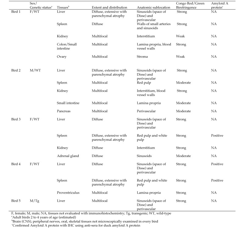
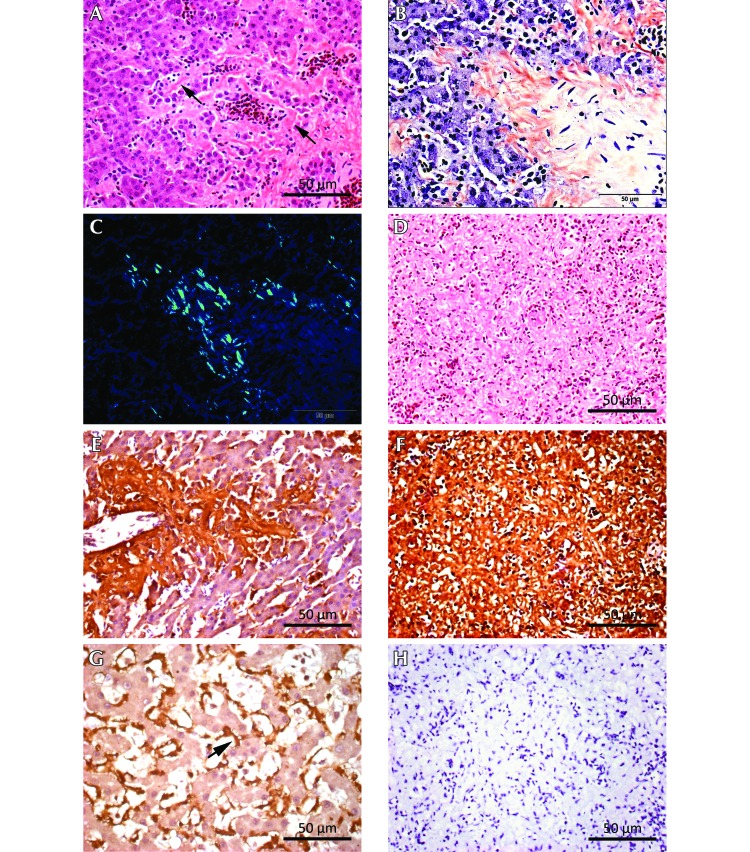
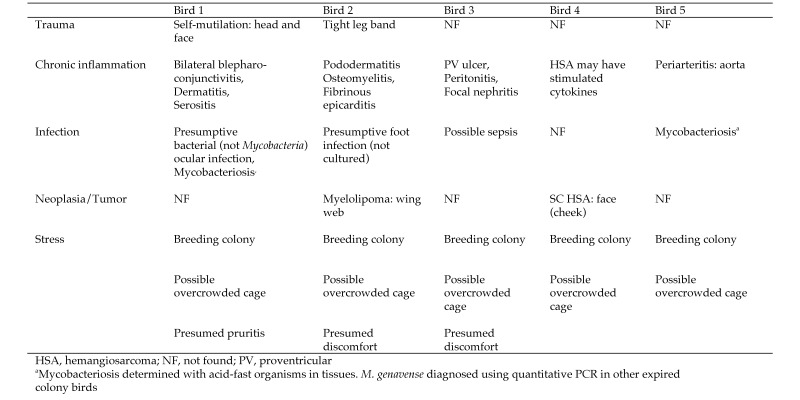
Five birds in a captive zebra finch research colony were diagnosed with systemic amyloidosis within a 7-mo period by means of postmortem Congo red staining and green birefringence under polarized light. The liver was the most frequently and usually the most seriously affected organ, followed by the spleen and then the kidney. All 5 birds had been clinically affected with various inflammatory, infectious, and neoplastic conditions associated with amyloid A (AA) amyloidosis in humans and animals. Immunohistochemistry using antisera against duck AA protein revealed that tissues from 2 of the 5 birds were positive for the presence of AA protein and systemic inflammation-associated amyloidosis. Although the development of AA amyloidosis has been associated with chronic inflammation, trauma, and various infectious and neoplastic diseases as well as possible genetic predispositions and stresses linked to overcrowding, the root causes for individual cases of AA amyloidosis are incompletely understood. As far as we know, this report is the first description of AA amyloidosis in captive, research zebra finches.


Figures

Figure 1.
Figure 1.
Amyloid lesions in birds 1 through 5.
Figure 2.
Figure 2.
Histopathology and immunohistochemstry for birds 3 and 4, with positive-control tissue from a Siamese cat. (A) Bird 3, liver. Diffuse, acellular eosinophilic material within the hepatic sinusoids (space of Disse, black arrows). Hematoxylin and eosin stain; scale bar, 50 µm. (B) Bird 3, liver. Salmon-stained areas (congophilia) correspond to diffuse sinusoidal amyloid deposits visible in the hematoxylin- and eosin-stained tissue shown in panel A. Congo red stain; scale bar, 50 µm. (C) Bird 3, liver. The Congo-red–stained section shows green birefringence (GBR), denoting amyloid protein, under polarized light. Scale bar, 50 µm. (D) Bird 3, spleen. Diffuse amyloid deposition involving both red and white pulp, with extensive loss of normal architecture. Hematoxylin and eosin stain; scale bar, 50 µm. (E) Bird 4, liver. Immunohistochemical localization of amyloid A (AA) protein shows copious brown reaction product within the hepatic sinusoids, with distribution similar to that of the amyloid detected by using congophilia and GBR. Antisera against duck AA protein, 1:600 dilution; scale bar, 50 µm. (F) Bird 3 spleen. Immunohistochemical localization of AA protein shows copious brown reaction product, with diffuse distribution similar to that of amyloid detected by using congophilia and GBR. Antisera against duck AA protein, 1:600 dilution; scale bar, 50 µm. (G) Siamese cat, liver. Positive control for immunohistochemical detection of AA protein shows brown reaction product (black arrow) within the sinusoids. Antisera against duck AA protein, 1:600 dilution; scale bar, 50 µm. (H) Bird 3, spleen. Omission of the incubation step from the staining procedure with primary antisera against duck AA protein shows no staining for AA protein. Scale bar, 50 µm.
Figure 3.
Figure 3.
Comorbidity classification system for birds with amyloidosis.

References

    1. Animal Welfare Act as Amended. 2008. 7 USC §2131–2156.
    1. Azzopardi JG, Lehner T. 1966. Systemic amyloidosis and malignant disease. J Clin Pathol 19:539–548. - PMC - PubMed
    1. Blancas-Mejia LM, Ramirez-Alvarado M. 2013. Systemic amyloidoses. Annu Rev Biochem 82:745–774. - PMC - PubMed
    1. Boyce JT, DiBartola SP, Chew DJ, Gasper PW. 1984. Familial renal amyloidosis in Abyssinian cats. Vet Pathol 21:33–38. - PubMed
    1. Buxbaum J. 2006. The genetics of the amyloidoses: interactions with immunity and inflammation. Genes Immun 7:439–449. - PubMed

Publication types

Substances