

A Supercomplex Spanning the Inner and Outer Membranes Mediates the Biogenesis of β-Barrel Outer Membrane Proteins in Bacteria
- PMID: 27298319
- PMCID: PMC4974385
- DOI: 10.1074/jbc.M115.710715
A Supercomplex Spanning the Inner and Outer Membranes Mediates the Biogenesis of β-Barrel Outer Membrane Proteins in Bacteria
Abstract
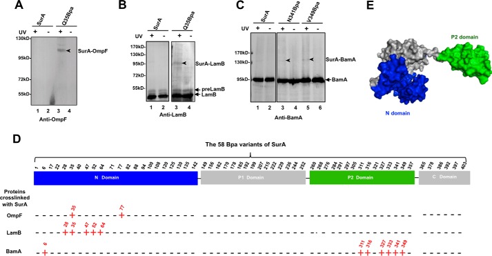
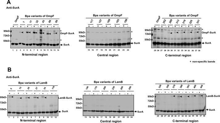
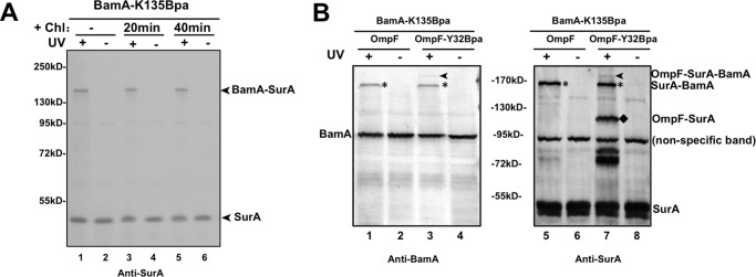
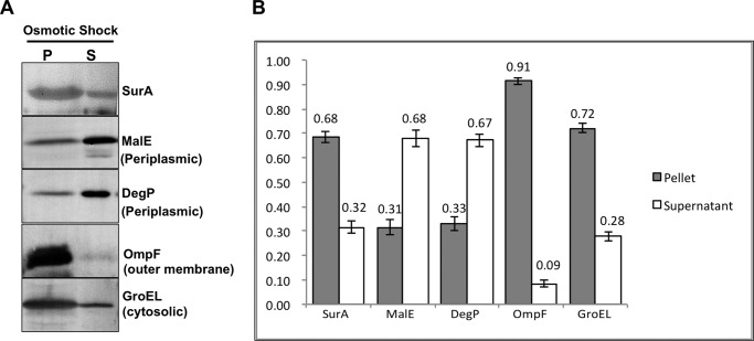
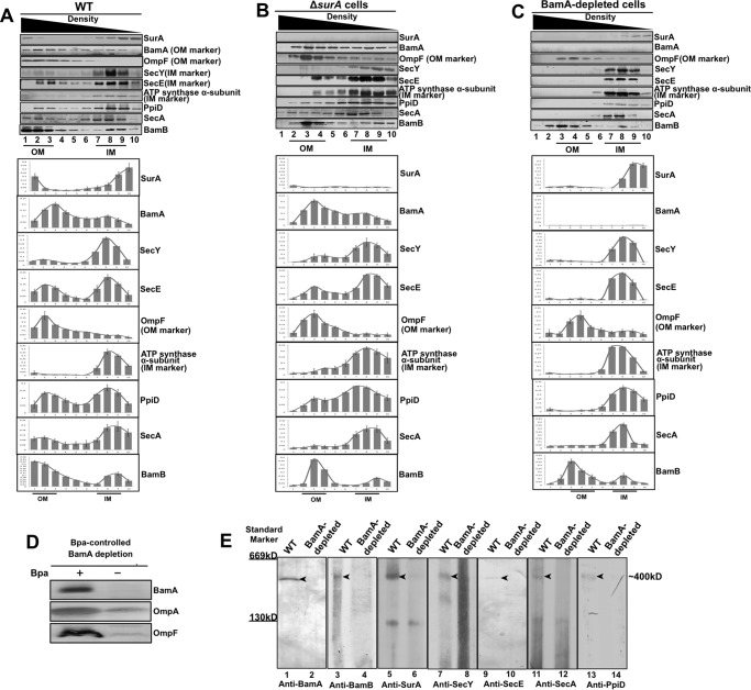
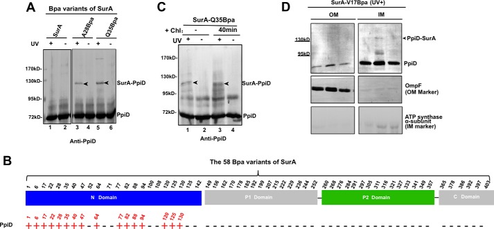
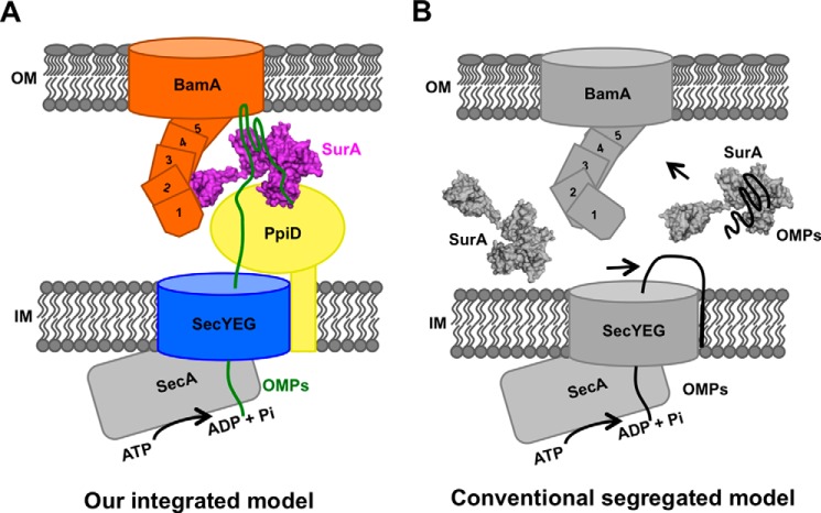
β-barrel outer membrane proteins (OMPs) are ubiquitously present in Gram-negative bacteria, mitochondria and chloroplasts, and function in a variety of biological processes. The mechanism by which the hydrophobic nascent β-barrel OMPs are transported through the hydrophilic periplasmic space in bacterial cells remains elusive. Here, mainly via unnatural amino acid-mediated in vivo photo-crosslinking studies, we revealed that the primary periplasmic chaperone SurA interacts with nascent β-barrel OMPs largely via its N-domain but with β-barrel assembly machine protein BamA mainly via its satellite P2 domain, and that the nascent β-barrel OMPs interact with SurA via their N- and C-terminal regions. Additionally, via dual in vivo photo-crosslinking, we demonstrated the formation of a ternary complex involving β-barrel OMP, SurA, and BamA in cells. More importantly, we found that a supercomplex spanning the inner and outer membranes and involving the BamA, BamB, SurA, PpiD, SecY, SecE, and SecA proteins appears to exist in living cells, as revealed by a combined analyses of sucrose-gradient ultra-centrifugation, Blue native PAGE and mass spectrometry. We propose that this supercomplex integrates the translocation, transportation, and membrane insertion events for β-barrel OMP biogenesis.
Keywords: BamA; SurA; chaperone; complex; membrane biogenesis; membrane protein; protein folding; the Sec translocon; β-barrel outer membrane protein biogenesis.
© 2016 by The American Society for Biochemistry and Molecular Biology, Inc.
Figures
Similar articles
-
The transmembrane supercomplex mediating the biogenesis of OMPs in Gram-negative bacteria assumes a circular conformational change upon activation.FEBS Open Bio. 2020 Aug;10(8):1698-1715. doi: 10.1002/2211-5463.12922. Epub 2020 Jul 23. FEBS Open Bio. 2020. PMID: 32602996 Free PMC article.
-
Substitutions in the BamA β-barrel domain overcome the conditional lethal phenotype of a ΔbamB ΔbamE strain of Escherichia coli.J Bacteriol. 2012 Jan;194(2):317-24. doi: 10.1128/JB.06192-11. Epub 2011 Oct 28. J Bacteriol. 2012. PMID: 22037403 Free PMC article.
-
Effects of Periplasmic Chaperones and Membrane Thickness on BamA-Catalyzed Outer-Membrane Protein Folding.J Mol Biol. 2017 Nov 24;429(23):3776-3792. doi: 10.1016/j.jmb.2017.09.008. Epub 2017 Sep 15. J Mol Biol. 2017. PMID: 28919234 Free PMC article.
-
Augmenting β-augmentation: structural basis of how BamB binds BamA and may support folding of outer membrane proteins.J Mol Biol. 2011 Mar 11;406(5):659-66. doi: 10.1016/j.jmb.2011.01.002. Epub 2011 Jan 12. J Mol Biol. 2011. PMID: 21236263 Review.
-
Building Better Barrels - β-barrel Biogenesis and Insertion in Bacteria and Mitochondria.J Mol Biol. 2021 Aug 6;433(16):166894. doi: 10.1016/j.jmb.2021.166894. Epub 2021 Feb 24. J Mol Biol. 2021. PMID: 33639212 Free PMC article. Review.
Cited by
-
The Escherichia coli Outer Membrane β-Barrel Assembly Machinery (BAM) Anchors the Peptidoglycan Layer by Spanning It with All Subunits.Int J Mol Sci. 2021 Feb 12;22(4):1853. doi: 10.3390/ijms22041853. Int J Mol Sci. 2021. PMID: 33673366 Free PMC article.
-
Pushing the Envelope: The Mysterious Journey Through the Bacterial Secretory Machinery, and Beyond.Front Microbiol. 2021 Nov 30;12:782900. doi: 10.3389/fmicb.2021.782900. eCollection 2021. Front Microbiol. 2021. PMID: 34917061 Free PMC article. Review.
-
Profiling the Escherichia coli membrane protein interactome captured in Peptidisc libraries.Elife. 2019 Jul 31;8:e46615. doi: 10.7554/eLife.46615. Elife. 2019. PMID: 31364989 Free PMC article.
-
Monitoring the Interaction of the Peptidoglycan with the Bacterial β-Barrel Assembly Machinery.Methods Mol Biol. 2024;2778:159-183. doi: 10.1007/978-1-0716-3734-0_11. Methods Mol Biol. 2024. PMID: 38478278
-
Similarly slow diffusion of BAM and SecYEG complexes in live E. coli cells observed with 3D spt-PALM.Biophys J. 2023 Nov 21;122(22):4382-4394. doi: 10.1016/j.bpj.2023.10.017. Epub 2023 Oct 17. Biophys J. 2023. PMID: 37853695 Free PMC article.
References
-
- Wimley W. C. (2003) The versatile β-barrel membrane protein. Curr. Opin. Struct. Biol. 13, 404–411 - PubMed
-
- Haltia T., and Freire E. (1995) Forces and factors that contribute to the structural stability of membrane-proteins. Biochim. Biophys. Acta Bioenergetics 1228, 1–27 - PubMed
-
- Koebnik R., Locher K. P., and Van Gelder P. (2000) Structure and function of bacterial outer membrane proteins: barrels in a nutshell. Mol. Microbiol. 37, 239–253 - PubMed
-
- Bos M. P., Robert V., and Tommassen J. (2007) Biogenesis of the gram-negative bacterial outer membrane. Annu. Rev. Microbiol. 61, 191–214 - PubMed
MeSH terms
Substances
Associated data
- Actions
- Actions
LinkOut - more resources
Full Text Sources
Other Literature Sources
Molecular Biology Databases
