. 2016 Jun 28;113(26):E3773-81.
doi: 10.1073/pnas.1604519113. Epub 2016 Jun 13.

Tau accumulation induces synaptic impairment and memory deficit by calcineurin-mediated inactivation of nuclear CaMKIV/CREB signaling

Affiliations

Tau accumulation induces synaptic impairment and memory deficit by calcineurin-mediated inactivation of nuclear CaMKIV/CREB signaling

Yaling Yin et al. Proc Natl Acad Sci U S A. .

Erratum in

Abstract

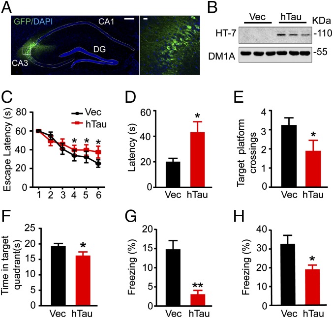
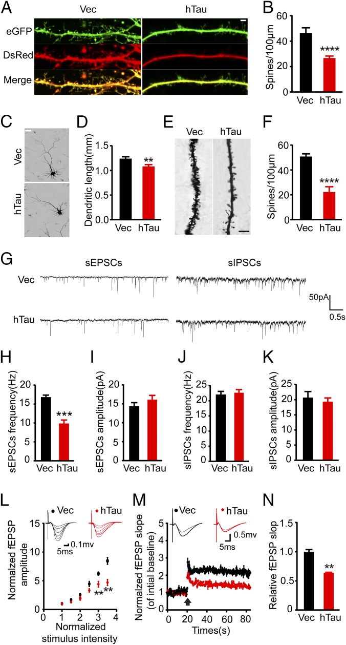
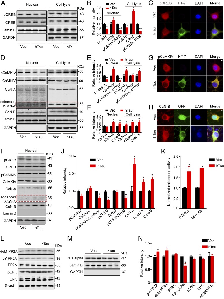
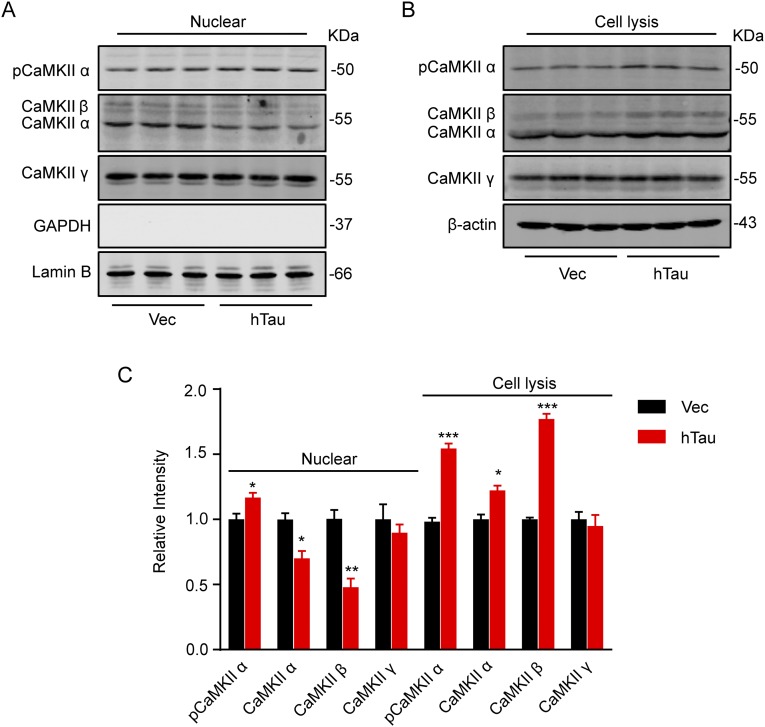
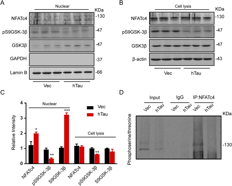
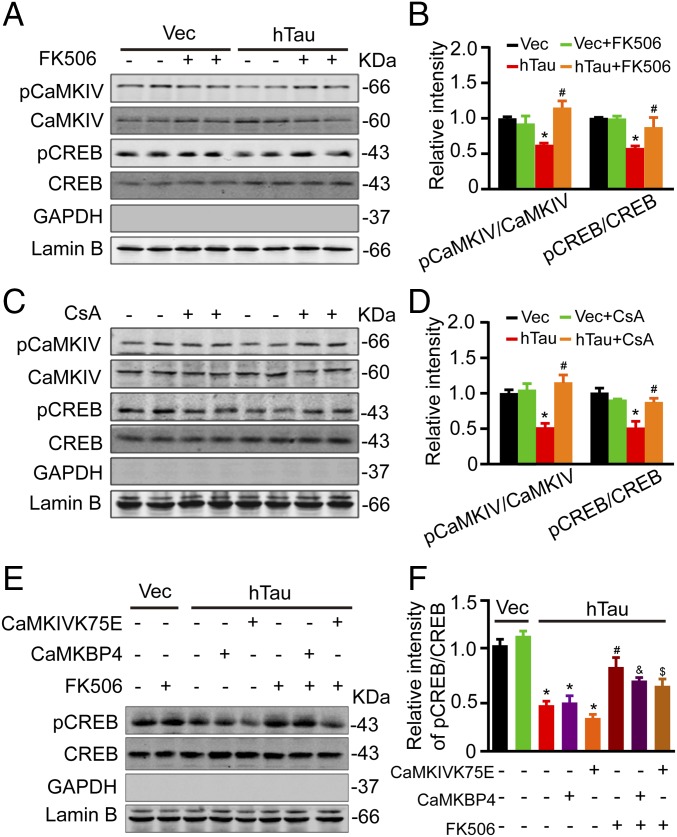
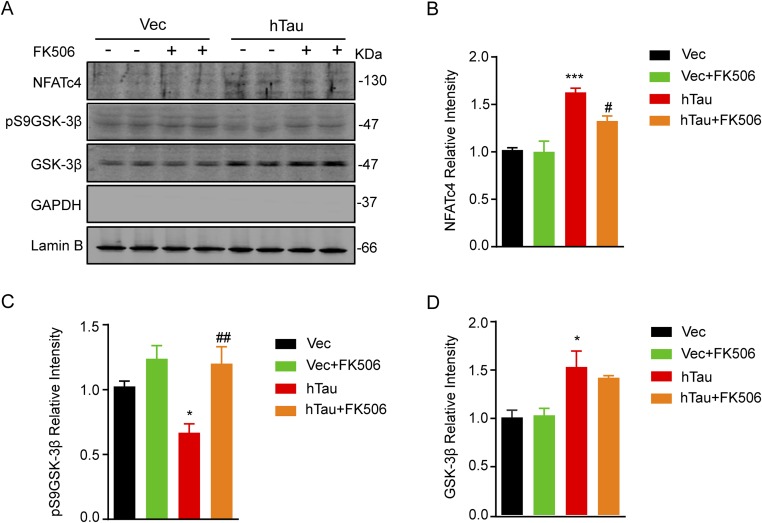
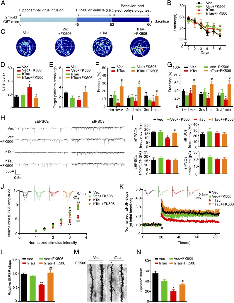
Intracellular accumulation of wild-type tau is a hallmark of sporadic Alzheimer's disease (AD), but the molecular mechanisms underlying tau-induced synapse impairment and memory deficit are poorly understood. Here we found that overexpression of human wild-type full-length tau (termed hTau) induced memory deficits with impairments of synaptic plasticity. Both in vivo and in vitro data demonstrated that hTau accumulation caused remarkable dephosphorylation of cAMP response element binding protein (CREB) in the nuclear fraction. Simultaneously, the calcium-dependent protein phosphatase calcineurin (CaN) was up-regulated, whereas the calcium/calmodulin-dependent protein kinase IV (CaMKIV) was suppressed. Further studies revealed that CaN activation could dephosphorylate CREB and CaMKIV, and the effect of CaN on CREB dephosphorylation was independent of CaMKIV inhibition. Finally, inhibition of CaN attenuated the hTau-induced CREB dephosphorylation with improved synapse and memory functions. Together, these data indicate that the hTau accumulation impairs synapse and memory by CaN-mediated suppression of nuclear CaMKIV/CREB signaling. Our findings not only reveal new mechanisms underlying the hTau-induced synaptic toxicity, but also provide potential targets for rescuing tauopathies.

Keywords: Alzheimer’s disease; CREB; Ca2+/calmodulin-dependent kinase IV; calcineurin; tau.


Conflict of interest statement

The authors declare no conflict of interest.

Figures

Fig. 1.
Fig. 1.
Tau accumulation in the hippocampal CA3 region impairs learning and memory. (A) The representative immunofluorescence imaging of hippocampus and the enlarged CA3 subset after infusion of AAV-eGFP-hTau for 6 wk. [Scale bars: 200 μm (Left) and 20 μm (Right).] (B) Expression of hTau in hippocampal CA3 was confirmed by Western blotting using human tau-specific antibody HT7. (C) The escape latency to find the hidden platform in MWM during 6-d learning process. (D–F) The escape latency to find the hidden platform, the target platform crossings, and the time spent in the target quadrant measured on day 8 by removing the platform. (G) After a 1-wk rest, fear conditioning was used to measure the contextual memory: the mice were exposed to foot shocks for 2 s (0.8 mA) followed by an auditory cue. After 24 h, the mice were put into the same training chamber without shocks and the auditory cue, and the total freezing time in 3 min was recorded with a video camera. (H) Two hours later, the freezing time in 3 min was measured again by putting the mice back into the same chamber with the auditory cue for 30 s. Data were expressed as mean ± SEM, *P < 0.05, **P < 0.01 vs. vector (Vec).
Fig. 2.
Fig. 2.
Overexpression of hTau decreases spine density and the dendrite length with impaired synapse transmission. (A) The representative images showing spine density in primary hippocampal neurons (12 div). cotransfection with DsRed and eGFP-hTau (hTau) or DsRed and eGFP (Vec) for 48 h, the images were visualized by using two-photon confocal laser scanning microscopy. (Scale bar, 2 μm.) (B) Quantitative analysis of the spine numbers (at least 30 neurons from three independent cultures were analyzed for each group). (C and D) The primary hippocampal neurons were probed by anti–MAP-2 and the dendrite length was analyzed by Image-pro plus, at least 30 neurons from three independent cultures were analyzed for each group). (Scale bar, 20 μm.) (E) The spine density in hippocampal CA3 with overexpression of hTau or the vector measured by Golgi stain. (Scale bar, 10 μm.) (F) Quantitative analysis of the spine numbers (at least 15 neurons, three dendritic branches per neuron, from three mice were used for the analysis). (G) The representative traces of sEPSCs and sIPSCs measured by whole-cell voltage patch-clamp recording on ex vivo brain slices. (H–K) Quantitative analyses of the sEPSC and sIPSC. (L) The IO curve of the fEPSP in CA3-CA1, normalized by fEPSP amplitude induced by minimum stimulation intensity. (M) The slope of fEPSP after HFS, normalized by the baseline. Arrow indicates the onset of HFS, the traces are average fEPSPs from five sweeps before (thin) and after (thick) LTP induction. (N) Quantitative analyses for fEPSPs measured 60–80 min after HFS relative to baseline. Data were expressed as mean ± SEM, *P < 0.05,**P < 0.01, ***P < 0.001, ****P < 0.0005 vs. Vec.
Fig. 3.
Fig. 3.
Overexpression of hTau inactivates nuclear CREB with up-regulation of CaN and inhibition of CaMKIV. (A and B) Hippocampal neurons (7 div) were infected with lenti-mCherry-hTau or the vector and cultured for 5 more days, then the cell lysates and the nuclear fraction were prepared for Western blotting of total and the Ser133-phosphorylated levels of CREB. (C) The representative immunofluorescence imaging showing reduced nuclear staining of pCREB (red) in neurons with overexpression of hTau probed by HT7 (green). (Scale bar, 5 μm.) (D–F) The protein levels of total and the pCaMKIV, total CaN-A and CaN-B, and the cleaved CaN-A (cCaN-A) in cell lysates and the nuclear fraction detected by Western blotting. (G and H) The reduced pCaMKIV (red) and increased CaN-B (red) in the nuclear fraction of the primary hippocampal neurons detected by immunofluorescent staining. (Scale bar, 5 μm.) (I and J) The increased protein levels of CaN-A and cCaN-A, CaN-B, and reduced pCaMKIV and pCREB detected in nuclear fraction of the hippocampal CA3 subset after infusion of AAV-eGFP-hTau for 6 wk. (K) The CaN activity in primary cultured hippocampal neurons (PCHNs) and mouse hippocampus CA3 (MHCA3) detected by using the activity assay kit. (LN) Total PP2A, the demethylated PP2A at Leu309 (deML309-PP2A), and the phosphorylated PP2A at tyrosine-307 (pY307-PP2A), total ERK, and pERK in cell lysates, and the level of PP1 catalytic subunit in the nuclear fraction measured by Western blotting. Lamin B and DAPI were used, respectively, as nuclear markers. Data were expressed as mean ± SEM, *P < 0.05, **P < 0.01 vs. Vec.
Fig. S1.
Fig. S1.
Effects of tau overexpression on CaMKII subunits. Hippocampal neurons (7 div) were infected with lenti-mCherry-hTau or the vector (Vec) and cultured for 5 more days; then the cell lysates and the nuclear fraction were prepared for Western blotting analyses of CaMKII subunits. The results showed that levels of total CaMKIIα, p-CaMKIIα and total CaMKIIβ were significantly increased, whereas CaMKIIγ was not changed in lysates (A and C); in the nuclear fraction, p-CaMKIIα significantly increased with reduced total CaMKIIα and CaMKIIβ and unchanged CaMKIIγ upon hTau overexpression (B and C). Data were presented as mean ± SEM, *P < 0.05, **P < 0.01, ***P < 0.001 vs. Vec.
Fig. S2.
Fig. S2.
Effects of tau overexpression on NFATc4 and GSK-3β phosphorylation. (AC) AAV-eGFP (Vec) or AAV-eGFP-hTau (hTau) was injected stereotaxically into the hippocampal CA3 of 2-mo-old mice. After 45 d, the hippocampal CA3 subset was dissected for Western blotting or immunoprecipitation analysis. The total NFATc4 level was not changed in lysate but it was significantly increased in the nuclear fraction, whereas the level of pS9GSK-3β was decreased in both lysate and the nuclear fractions. (D) NFATc4 in the hippocampal CA3 extract was immunoprecipitated by using anti-NFATc4, and the phosphorylation of NFATc4 was detected by using antiphosphoserine/threonine antibody. A reduced NFAT4c phosphorylation at serine and threonine residuals was shown after hTau expression. Data were represented as mean ± SEM, *P < 0.05, ***P < 0.001 vs. Vec.
Fig. 4.
Fig. 4.
Inhibition of CaN attenuates the hTau-induced CREB dephosphorylation independent of its effects on CaMKIV inhibition. (A–D) The primary hippocampal neurons (7 div) were infected with lenti-mCherry-hTau or the vector, after being cultured for 5 more days, the neurons were treated with 1 μM FK506 or 50 nM CsA for 12 h, then the levels pS133-CREB and pS196-CaMKIV were measured by Western blotting. (E and F) N2A cells, cotransfected with hTau plus CaMKBP4 or CaMKIVK75E plasmids for 48 h, were treated with 1 μM FK506 for 12 h, and then the phosphorylation level of CREB in the nuclear fraction was measured by Western blotting. Data were expressed as mean ± SEM, *P < 0.05 vs. Vec; #P < 0.05 vs. hTau; and &P < 0.05 vs. hTau plus CaMKBP4; $P < 0.05 vs. hTau plus CaMKIVK75E.
Fig. S3.
Fig. S3.
Simultaneous inhibition of CaN by FK506 attenuates the hTau-induced GSK-3β and NFATc4 dephosphorylation. AAV-eGFP (Vec) or AAV-eGFP-hTau (hTau) was injected stereotaxically into the hippocampal CA3 of 2-mo-old mice. After 45 d, FK506 (10 mg⋅kg⋅d) or the vehicle was injected intraperitoneally for 1 wk. Then the hippocampal CA3 subset was dissected for Western blotting (A). Quantitative analysis showed that simultaneous inhibition of CaN increased the pS9GSK-3β level (C) with an increased total GSK-3β (D) and a slight decrease of NFATc4 (B) in the nuclear fraction. Data were represented as mean ± SEM, *P < 0.05, ***P < 0.001 vs. Vec; #P < 0.05, ##P < 0.01 vs. hTau.
Fig. 5.
Fig. 5.
Inhibition of CaN rescues the hTau-induced memory deficits with improvement of dendritic plasticity and synaptic functions. (A) Schematics show the treatments. AAV-eGFP (Vec) or AAV-eGFP-hTau (hTau) was injected stereotaxically into the hippocampal CA3 of 2-mo-old mice. After 45 d, FK506 (10 mg⋅kg⋅d) or the vehicle was injected intraperitoneally for 1 wk. Then the cognitive behaviors and synaptic plasticity were detected. (B) The escape latency to find the hidden platform during the 6-d learning process in MWM test. (C) The representative swimming tracks during the memory test carried out on day 8 by removing the hidden platform. (D and E) The escape latency to find the platform and target platform crossings tested on day 8. (F and G) Fear conditioning was used to measure the contextual memory of the mice 1 wk after the last MWM task. (H) The representative traces of sEPSCs and sIPSCs recorded by whole-cell voltage patch-clamp on ex vivo brain slices after FK506 or vehicle treatment. (I) The average frequency and amplitude of sEPSCs or sIPSCs collected from at least 12 neurons per group. (Scale bars, 10 pA, 1 s.) (J) The IO curve of fEPSP recorded on acute hippocampal slices overexpressing hTau or the vector and treated with FK506 or the vehicle (n = 6 per group). (K) The slope of fEPSP after HFS recorded on hippocampal slices after FK506 or vehicle treatment (n = 6 per group). Arrow indicates HFS onset, the average traces fEPSPs before (thin) and after (thick) LTP induction are shown. (L) Quantitative analyses for normalized fEPSPs 60–80 min after HFS. (M and N) The spine density in hippocampal CA3 subset imaged by Golgi staining. The data were expressed as mean ± SEM, *P < 0.05, **P < 0.01, Vec vs. hTau; #P < 0.05, ##P < 0.01, hTau vs. hTau plus FK506.

References

    1. Bennett DA, Schneider JA, Bienias JL, Evans DA, Wilson RS. Mild cognitive impairment is related to Alzheimer disease pathology and cerebral infarctions. Neurology. 2005;64(5):834–841. - PubMed
    1. Glenner GG, Wong CW. Alzheimer’s disease: Initial report of the purification and characterization of a novel cerebrovascular amyloid protein. Biochem Biophys Res Commun. 1984;120(3):885–890. - PubMed
    1. Grundke-Iqbal I, et al. Microtubule-associated protein tau. A component of Alzheimer paired helical filaments. J Biol Chem. 1986;261(13):6084–6089. - PubMed
    1. Davies CA, Mann DM, Sumpter PQ, Yates PO. A quantitative morphometric analysis of the neuronal and synaptic content of the frontal and temporal cortex in patients with Alzheimer’s disease. J Neurol Sci. 1987;78(2):151–164. - PubMed
    1. DeKosky ST, Scheff SW. Synapse loss in frontal cortex biopsies in Alzheimer’s disease: Correlation with cognitive severity. Ann Neurol. 1990;27(5):457–464. - PubMed

Publication types

MeSH terms

Substances

LinkOut - more resources