. 2016 Jun 21;113(25):6916-21.
doi: 10.1073/pnas.1603544113. Epub 2016 Jun 13.

δ-COP contains a helix C-terminal to its longin domain key to COPI dynamics and function

Affiliations

δ-COP contains a helix C-terminal to its longin domain key to COPI dynamics and function

Eric C Arakel et al. Proc Natl Acad Sci U S A. .

Abstract

Membrane recruitment of coatomer and formation of coat protein I (COPI)-coated vesicles is crucial to homeostasis in the early secretory pathway. The conformational dynamics of COPI during cargo capture and vesicle formation is incompletely understood. By scanning the length of δ-COP via functional complementation in yeast, we dissect the domains of the δ-COP subunit. We show that the μ-homology domain is dispensable for COPI function in the early secretory pathway, whereas the N-terminal longin domain is essential. We map a previously uncharacterized helix, C-terminal to the longin domain, that is specifically required for the retrieval of HDEL-bearing endoplasmic reticulum-luminal residents. It is positionally analogous to an unstructured linker that becomes helical and membrane-facing in the open form of the AP2 clathrin adaptor complex. Based on the amphipathic nature of the critical helix it may probe the membrane for lipid packing defects or mediate interaction with cargo and thus contribute to stabilizing membrane-associated coatomer.

Keywords: ARCN1; COPI; HDEL; KDEL; coatomer.


Conflict of interest statement

The authors declare no conflict of interest.

Figures
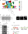
Fig. S1.
Fig. S1.
The B. taurus δ-COP subunit ARCN1 functionally complements the essential yeast δ-COP subunit RET2 despite sharing only 34% sequence similarity. (A) Schematic illustration of membrane-associated coatomer based on ref. . The Ret2 subunit of the yeast COPI coat was replaced with the Archain subunit of B. taurus in the δc* strain. (B) Binding of coatomer from yeast cytosol to GST fusion proteins of the cytosolic tail of Mst27 presenting the indicated di-lysine (-KKXX) or Arg-based (-ΦRXR) ER retrieval signals (14). Cytosol was prepared from a wild-type BY4741 strain or the δc* strain. The bound fraction was eluted from the affinity matrix and analyzed by SDS/PAGE followed by immunoblot analysis using an antiserum recognizing all seven coatomer subunits. Five percent of the input cytosol was loaded as a control. (C) Localization analysis of a Pmp2YFP reporter protein presenting a di-lysine (-KKXX), an Arg-based (-ΦRXR), or a weak (NVRNRRK) Arg-based ER retrieval signal (14) in the δC* strain. (D) TAP purification of coatomer from a Sec27-TAP (β′-COP) tagged wild-type BY4741 or δc* strain. The eluted coat was analyzed by SDS/PAGE followed by immunoblot analysis using an antiserum recognizing all seven coatomer subunits and antibodies specific for Ret2 and δ-COP. Note that the signal intensity of the trunk/ F-sub complex (β, γ) in the δc* strain is significantly lower than that of the wild-type BY4741 strain, presumably owing to the dissociation of the trunk during purification. The signal intensity of the cage/ B-subcomplex is identical in both BY4741 and δc* strains. (E) The boundaries at which the chimaeras were constructed, mapped onto the predicted secondary structure of Ret2. The longin domain (LD) and the μHD are indicated.
Fig. 1.
Fig. 1.
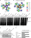
Expansion on an incomplete COPI structure via functional dissection of a highly flexible coatomer subunit. (A) Secretion assay of a ret2 deletion strain harboring a functional B. taurus ARCN1 gene (δc*) was transformed with plasmids containing Ret2-Archain chimaeras (CHI 1–8). Proteins secreted into the culture medium (i.e., extracellular) were analyzed by SDS/PAGE and immunoblot analysis using antibodies specific for Kar2 and Pdi1. Cell pellets from the same cultures were also similarly analyzed using antibodies specific for Pgk1. (B) Schematic representation of the Ret2-Archain chimaeras used in A. Boundaries at which the chimaeras were constructed and alignments of their sequences are indicated. (C) Chimaera 2, containing the longin domain and two predicted adjoining helices of Ret2 rescues the Pdi1 secretion phenotype of δc*. Bar graph of the relative amounts of Pdi1 secreted into the culture medium as quantified by the densitometric analysis of immunoblots. Quantification of five independent experiments. Error bars depict SEM.
Fig. S2.
Fig. S2.
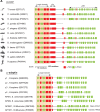
Prediction-based structural alignment of δ-COP and μ-adaptin reveals minor but important variations in its architecture. (A) Schematic representation of JPred secondary structure predictions of the δ-COP subunit from different species. The longin domain, the two adjoining helices (+2α), and the μHD are conserved from simple eukaryotes such as yeast to higher eukaryotes such as mammals. Note that S. pombe lacks a functional μHD. The UniProt accession numbers have been provided in parentheses. (B) Schematic representation of JPred secondary structure predictions of the μ-adaptin subunits from different species. The JPred secondary structure predictions indicate significant structural similarity between the μ-adaptin and the δ-COP subunit (A), apart from the second helix C-terminal to the longin domain. The UniProt accession numbers have been provided in parentheses.
Fig. 2.
Fig. 2.
The μHD of Ret2 is dispensable. (A) Schematic representation of JPred secondary structure predictions of the δ-COP subunit from different species. Note that S. pombe lacks a functional μHD. The secondary structure of μ-adaptin has been illustrated for comparison. The UniProt accession numbers have been provided within parentheses. (B) Growth assay demonstrating that deletion of the Ret2 μHD does not cause temperature sensitivity up to 39 °C. (C) Secretion analysis of ret2 deletion strains expressing ARCN1, RET2, or truncated RET2 (i.e., lacking the μHD; Ret2LD2α). Proteins secreted into the culture medium were analyzed by SDS/PAGE and immunoblot analysis using antibodies specific for Pdi1 and Kar2. Cell pellets from the same cultures were similarly analyzed using antibodies specific for Pgk1, coatomer, and a His-tag present on Ret2LD2α. Note that the coat antibody detects the μHD of Ret2.
Fig. 3.
Fig. 3.
A predicted helix C-terminal to the Ret2 longin domain plays a pivotal role in the HDEL-dependent retrieval system. (A) Schematic representation of the JPred secondary structure predictions of the truncated and chimeric constructs of Ret2 and Archain used in B. α-Helices are colored red and purple and β-sheets are green and brown (for Ret2 and Archain, respectively). (B) ret2 deletion strains containing either truncated genes or chimaeras of RET2 and ARCN1 (i.e., lacking the μHD, the μHD and the two helices C-terminal to the longin domain and chimaeras with swapped helices). Proteins secreted into the culture medium were analyzed by SDS/PAGE and immunoblot analysis using antibodies specific for Pdi1. Cell pellets from the same cultures were also similarly analyzed using antibodies specific for Ret2, Archain, coatomer, and Pgk1. (C) Bar graph of the relative amounts of Pdi1 secreted into the culture medium as quantified by the densitometric analysis of immunoblots. Quantification of three independent experiments. Error bars depict SEM.
Fig. 4.
Fig. 4.
Seventeen residues at the core of the identified α-helix are key to the retrieval of HDEL-bearing cargo. (A) Schematic representation of JPred secondary structure predictions. α-Helices are colored red and purple and β-sheets are green and brown (for Ret2 and Archain, respectively). Nomenclature of longin domain secondary structural elements corresponds to ref. . (B) ret2 deletion strains containing chimeric constructs of Ret2 and Archain in the two helices C-terminal to the longin domain were analyzed for the secretion of HDEL-bearing proteins. Proteins secreted into the culture medium were analyzed by SDS/PAGE and immunoblot analysis using antibodies specific for Pdi1. Cell pellets from the same cultures were also similarly analyzed using antibodies specific for coat, Pgk1, and a His-tag present in Ret2LD2α. (C) Bar graph of the relative amounts of Pdi1 secreted into the culture medium as quantified by the densitometric analysis of immunoblots. Quantification of four independent experiments. Error bars depict SEM. (D) TAP-tagged coatomer purified from the three indicated strains. (E) Schematic illustration of the conformational change in COPI following membrane recruitment. Like AP2, this potentially results in displacement of the μHD of δ-COP, the opening of the coatomer core via extension of the F-subcomplex, and the binding of the newly identified helix in δ-COP (colored red) onto β-COP. The interaction of individual COPI subunits with cargo (through ER retrieval signals) further stabilizes the open conformation of coatomer. ArfGAP1 (colored purple) with a δL-motif interacts with the μHD of δ-COP and Arf1.
Fig. S3.
Fig. S3.
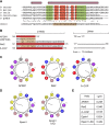
The identified helix has characteristics of an amphipathic helix. (A) Sequence alignment of the two predicted helices C-terminal to the δ-COP longin domain from different species. The highly conserved regions of the second helix have been highlighted in green. Chimaera 10 (CHI10) was created by replacing the 17 residues within the conserved regions of Ret2 with the corresponding residues of B. taurus Archain. Unconserved residues in this minimal region have been highlighted in red. (B) Amino acid sequence and length of the critical helix of μ-adaptin, Ret2, and δ-COP (Left). The residue positions are indicated. Schematic representation of the linker lengths connecting the critical helix and the μHD in μ-adaptin, Ret2 and δ-COP (Right). (C) Helical wheel representation of the helices of μ-adaptin, Ret2, and δ-COP. Residues have been color-coded: yellow for hydrophobic, purple for serine and threonine, blue for basic, red for acidic, pink for asparagine and glutamine, and gray for alanine and glycine. The helical wheel corresponds to the region highlighted in orange in B. The arrow in the helical wheel corresponds to the hydrophobic moment and its length reflects the size of the hydrophobic moment. (D) Helical wheel representation of the α0 helix of Epsin and mutant (L6Q) after ref. . The residues are color-coded as in C. (E) Table summarizing the hydrophobic moment (<μH>) obtained from helical wheel analysis using HeliQuest (40).
Fig. S4.
Fig. S4.
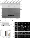
Mutations in the identified helix do not impinge on the canonical retrieval functions of other COPI subunits. (A) Binding of TAP-purified coatomer to GST fusion proteins of the cytosolic tail of Mst27 presenting the indicated di-lysine (-KKXX) or Arg-based (-ΦRXR) ER retrieval signals. Coat was purified from a wild-type BY4741 (C1) strain or strains expressing a Ret2LD2α (C2), CHI10 (C3), and Ret2LD (C4) variant of coat. The bound fraction was eluted from the affinity matrix and analyzed by SDS/PAGE followed by immunoblot analysis using an antiserum recognizing all seven coatomer subunits. C2, C3, and C4 contain a truncated Ret2 subunit, lacking the μHD that the coat antiserum detects. The purified coats were loaded as an input control (B). (C) Bar graph of the relative amounts of bound COPI as quantified by the densitometric analysis of three independent immunoblots similar to that depicted in A. The signal intensity of eluted COPI was normalized to the signal intensity of input COPI to correct for differences in the amount of coat used in each case. Error bars depict SEM. (D) Localization analysis of a Pmp2YFP reporter protein presenting a di-lysine (-KKXX), an Arg-based ER retrieval signal (-ΦRXR), its inactive mutants (-AAXX and -ΦKKK, respectively), or the C-terminus of the TASK3 ion channel (T3-CT) with a COPI binding signal [-KRRKSV (26)] in a wild-type strain, or a ret2 deletion strain expressing wild-type Ret2 or variants Ret2LD2α, CHI10, or Ret2LD.
Fig. S5.
Fig. S5.
Other canonical retrieval functions of COPI are unperturbed in cells with mutant coatomer, supporting a specific defect in the HDEL-based retrieval system. (A) Binding of TAP-purified coatomer to GST fusion proteins of the cytosolic tail of Mst27 or the Gcn4 coiled-coil forming domain presenting the di-lysine (-KXKXX) ER retrieval signals of Erp1 and a control, Erp2. Coat was purified from a wild-type BY4741 strain or strains containing a Ret2LD2α and CHI10 variant of coat. The bound fraction was eluted from the affinity matrix and analyzed by SDS/PAGE followed by immunoblot analysis using an antiserum recognizing all seven coatomer subunits. (B) Bar graph of the relative amounts of eluted/bound COPI as quantified by the densiometric analysis of three independent immunoblots similar to that depicted in A. The signal intensity of eluted COPI was normalized to the signal intensity of input COPI to correct for differences in the amount of coat used in each case. Error bars depict SEM.
Fig. S6.
Fig. S6.
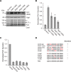
Three isoleucines within the identified helix are critical for its function. (A) Mutation of the hydrophobic residues within the critical stretch of the second helix C-terminal to the longin domain results in the secretion of HDEL-bearing proteins. A ret2 deletion strain was rescued with plasmids containing the indicated Ret2 variants. Proteins secreted into the culture medium (i.e., extracellular) were concentrated and harvested by TCA precipitation and analyzed by SDS/PAGE, followed by immunoblot analysis using antibodies specific for Pdi1. Cell pellets from the same cultures were also similarly analyzed using antibodies specific for Pgk1 and COPI. (B) Bar graph of the relative amounts of Pdi1 secreted into the culture medium as quantified by densitometric analysis of immunoblots similar to that depicted in A. Quantification of six independent experiments. Error bars depict SEM. (C) Same analysis as in A and B for constructs with individual residues within the critical helix changed from the residue present in Ret2 to that present in bovine δ-COP. Quantification of four independent experiments. Error bars depict SEM. (D) Qualitative summary of results obtained in B and C.
Fig. S7.
Fig. S7.
Linkages mediated by the ε-COP subunit or δ-COP μHD of coatomer are nonessential. Schematic illustration of the basic building block of the COPI coat, a triad linkage of coatomers, based on the structural model proposed by Dodonova et al. (3). Coatomer triads are thought to be linked by sets of interactions involving flexible domains such as ε-COP and the C-terminal domain of α-COP (Linkage I) (A) or via the μHD of δ-COP (Linkage III) (B). Note that the δ-COP μHD is either at the periphery or at the core of the triad depending on the type of linkage. These 2D schematic representations build on the crystal structures of PDB ID codes 5A1V and 5A1X, respectively. Unresolved domains such as the μHD in linkage I and αc and ε in linkage III are rendered translucently. (C) Growth assay demonstrating that the combined deletion of the Ret2 μHD and the ε-COP subunit does not affect viability up to 34 °C. The indicated yeast strains were grown to stationary phase, washed, and normalized to OD600 and serial dilutions were spotted onto YPAD agar and incubated at 25 °C, 30 °C, 34 °C, or 37 °C for 2 d. (D) SDS/PAGE and Western blot analysis of the indicated strains using antibodies specific for coat and Pgk1. (E) Bar graph of the relative amounts of α-COP in the indicated strains, as quantified by the densiometric analysis of three independent immunoblots similar to that depicted in D. Error bars depict SEM. Note that simultaneous deletion of the μHD neither suppresses nor exacerbates the lower steady-state levels of α-COP observed in the sec28 deletion strain. This indicates that deletion of the μHD does not further destabilize the coat and excludes the hypothesis that one type of linkage may compensate for the loss of the other.
Fig. S8.
Fig. S8.
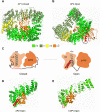
By analogy to AP2, the identified region may serve as a pivot around which δ-COP is structurally reorganized upon membrane recruitment of COPI. The AP2 adaptor complex undergoes a large-scale conformational change upon membrane recruitment, transitioning from a closed form (A) to an open structure (B). This change results in the relocation of the two large adaptor subunits, the C-terminal μ-adaptin subunit (which becomes coplanar with the membrane) and the transformation of an unstructured μ-adaptin linker to a helix (colored in red), which then binds back onto the complex. This structural rearrangement exposes previously occluded cargo and lipid binding sites. A and B correspond to the crystal structure of PDB ID codes 1W63 and 2XA7 (11, 41), respectively. (C) Schematic representation of the structural rearrangement upon membrane association where the unstructured linker transforms into an α-helix in the open conformation. The longin domain (LD), the second α-helix of the LD (αx), and the μHD of μ-adaptin have been highlighted for orientation. Comparison of the crystal structure of the β2-μ2-adaptin dimer (D) and the cryo-EM tomography derived β-δ-COP dimer (E). The β-δ-COP dimer appears as a ‟hyperopen” form compared with the β2-μ2-adaptin dimer. Note that the identified helix (corresponding to the second helix C-terminal to the longin domain) is absent in the homology model (marked with a red dotted ellipse).

References

    1. Pavel J, Harter C, Wieland FT. Reversible dissociation of coatomer: Functional characterization of a beta/delta-coat protein subcomplex. Proc Natl Acad Sci USA. 1998;95(5):2140–2145. - PMC - PubMed
    1. Lowe M, Kreis TE. In vitro assembly and disassembly of coatomer. J Biol Chem. 1995;270(52):31364–31371. - PubMed
    1. Dodonova SO, et al. VESICULAR TRANSPORT. A structure of the COPI coat and the role of coat proteins in membrane vesicle assembly. Science. 2015;349(6244):195–198. - PubMed
    1. Lee C, Goldberg J. Structure of coatomer cage proteins and the relationship among COPI, COPII, and clathrin vesicle coats. Cell. 2010;142(1):123–132. - PMC - PubMed
    1. Hsia KC, Hoelz A. Crystal structure of α-COP in complex with ϵ-COP provides insight into the architecture of the COPI vesicular coat. Proc Natl Acad Sci USA. 2010;107(25):11271–11276. - PMC - PubMed

Publication types