

Intramuscular Myxoma - A Rare Tumor
- PMID: 27298930
- PMCID: PMC4719287
- DOI: 10.13107/jocr.2250-0685.130
Intramuscular Myxoma - A Rare Tumor
Abstract
Introduction: Intramuscular myxoma is a rare benign soft tissue tumor involving the musculoskeletal system. The incidence is reported as varying from 0.1 to 0.13 per 100,000 population. Most patients present between the fifth and sixth decade of life. The swelling commonly occurs in the large muscles of the thigh, shoulder, buttocks and arms. The tumor can be diagnosed with certainty only with histopathological examination. Local recurrence is rare after excision.
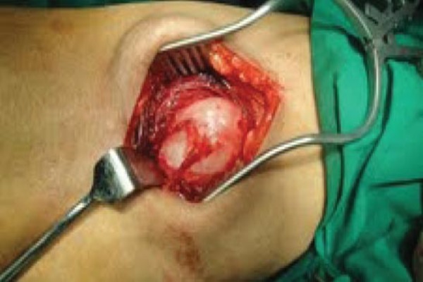
Case report: A 73 year old male patient presented to us with a swelling of the anterior aspect of the middle third of the right thigh measuring about 8 × 4 centimeters. He was thoroughly investigated and the swelling excised in toto. The soft cystic swelling excised was found on histopathology to be an intramuscular myxoma.
Conclusion: This case report is being presented since the tumor is rare and also an important consideration in the differential diagnosis of sarcomas, intramuscular lipoma, hemangioma, hematoma, and desmoid tumor. Another important feature is that it can be diagnosed with certainty only after excision.
Keywords: Intramuscular myxoma; benign; soft tissue swelling.
Conflict of interest statement
Conflict of Interest: Nil
Figures


References
-
- Tan HM, Peh WC, Shek TW. A distinctive shoulder mass. Br J Radiol. 2001;74:1159–1160. - PubMed
-
- Kabukcuoglu F, Kabukcuoglu Y. Mazabraud’s syndrome: Intramuscular myxoma associated with fibrous dysplasia. Orphanet Encyclopedia. 2005 Jan - PubMed
-
- Aind R A, Dwivedi M K, Pal R, Devangan L, Shekhar P V. Intra-muscular myxoma of gluteal region. Indian J Radiol Imaging. 2004;14:177–8.
-
- Ozbek N, Danaci M, Okumus B, Gursel B, Cakir S, Dabak N, Karagoz F. Recurrent intramuscular myxoma: review of the literature, diagnosis and treatment options. Turk J Cancer. 2006;36(2):75.
-
- Robert J Logel. Recurrent Intramuscular myxoma associated with Albright’s Syndrome. Journal of Bone and Joint Surgery. 58-A(4):565–568. - PubMed
Publication types
LinkOut - more resources
Full Text Sources
Other Literature Sources