

Congenital Pseudoarthrosis of Medial Malleolus in A Young Soccer Player - Diagnosis in Clinical setting of Ankle Sprain
- PMID: 27298936
- PMCID: PMC4722558
- DOI: 10.13107/jocr.2250-0685.139
Congenital Pseudoarthrosis of Medial Malleolus in A Young Soccer Player - Diagnosis in Clinical setting of Ankle Sprain
Abstract
Introduction: We report a case of a young female soccer player affected by congenital medial bilateral malleolus pseudoarthrosis and os subfibulare. Congenital pseudoarthrosis is the failure of the bones to fuse prior or at birth. The etiology is still unknown, although frequency is high in subjects affected by neurofibromatosis or correlated syndromes, so it has been suggested that these congenital disorders may be the cause of congenital pseudoarthrosis.
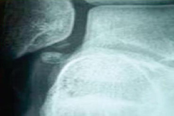
Case report: Our patient, a 16-year-old female, high level soccer player, was referred to us following a right ankle sprain during a match. She reported no medical history of tibia-tarsus joint injuries or disease. Pain, swelling and functional impairment were noted immediately after the accident. Standard radiographs in the emergency department revealed a displaced fracture of the medial malleolus and the presence of os subfibularis. The patient was transferred to our Traumatology and Orthopaedic Department to undergo malleolus ostheosynthesis. Before surgery swelling, functional impairment and intense pain at the medial malleolus level were confirmed. However, there was no radiological opening of ankle, instability or pronation pain; furthermore the flexion-extension was preserved with slight pain. Twenty-four hours later a considerable remission of symptoms was evident with increased range of motion and reduction in the swelling and post-traumatic edema. A radiograph on the left ankle to compare with that of the right ankle was necessary to overcome the discrepancy between the radiological diagnosis and the clinical examination. The radiographic results of both medial malleoli were comparable although on the left the os subfibularis was absent. Since the diagnosis of fracture by the association between the radiographs and the symptomatology was doubtful, a bilateral CT was performed. The scan revealed a medial bilateral malleolus pseudoarthrosis and an accessory right subfibularis nucleus. The patient was discharged from hospital with the diagnosis of "second degree right ankle sprain in patient affected by congenital medial bilateral malleolus pseudoarthrosis". A therapeutic- rehabilitative program was prescribed for the ankle sprain and unnecessary surgery was avoided. After 30 days there was an almost complete remission of pain. At a follow-up of six months the patient was completely asymptomatic and gradually began competitive activity.
Conclusion: An accurate history and an objective examination should be performed and correlated with the results of diagnostic procedures in order to avoid the incorrect diagnosis of a fracture needing surgery. The rarity of this ailment and the absence of consequences on long-term function, show that this disease does not justify sports activity cessation. Traumatic events at this site must be assessed properly in order to avoid being confused with malleolus fractures leading to over treatment.
Keywords: Ankle sprain; medial malleolus; pseudoarthrosis Introduction.
Conflict of interest statement
Conflict of Interest: Nil
Figures


References
-
- Harris NH. Massachusetts, U.S.A: John Wright & Soons Ltd; 1983. Postgraduate textbook of clinical orthopaedics.
-
- Canale ST, Beaty JH. 2nd ed. St. Louis: Mosby-Year Book; 1995. Operative Pediatric Orthopaedics.
-
- Delgado-Martinez AD, Rodriguez-Merchan EC, Olsen B. Congenital pseudarthrosis of the tibia. Int Orthop. 1996;20:192–199. - PubMed
-
- Hefti F, Bollini G, Dungl P, et al. Congenital pseudarthrosis of the tibia: history, etiology, classification, and epidemiologic data. J Pediatr Orthop B. 2000;9:11–15. - PubMed
-
- Ogden JA, Ganey TM, Ogden DA. The histopathology of injury to the accessory malleolar ossification center. J Pediatr Orthop. 1996;16:61–62. - PubMed
Publication types
LinkOut - more resources
Full Text Sources
Other Literature Sources
Research Materials