

"Soap Bubble" Lesion of the Middle Phalanx: Enchondroma or Epitheloid Hemangioma
- PMID: 27298959
- PMCID: PMC4719374
- DOI: 10.13107/jocr.2250-0685.167
"Soap Bubble" Lesion of the Middle Phalanx: Enchondroma or Epitheloid Hemangioma
Abstract
Introduction: Epitheloid hemangioma, a benign vascular tumor that arises in skin and soft tissues can also involve the skeletal system. Occasionally this has been reported from small tubular bones of the hand.
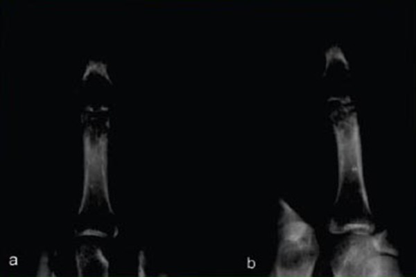
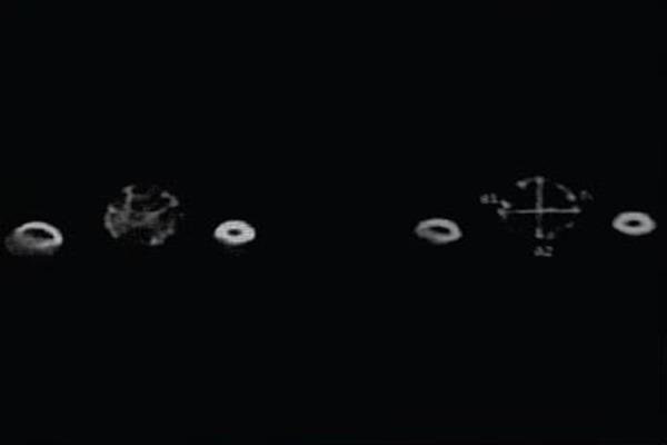
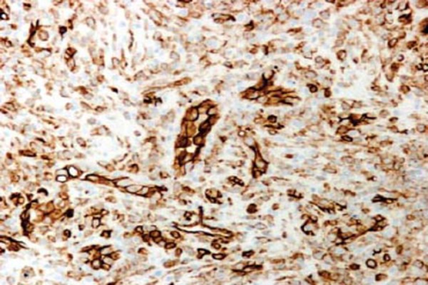
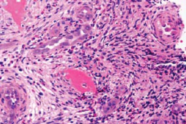
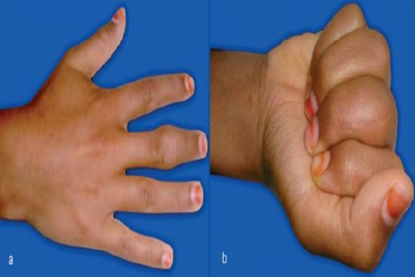
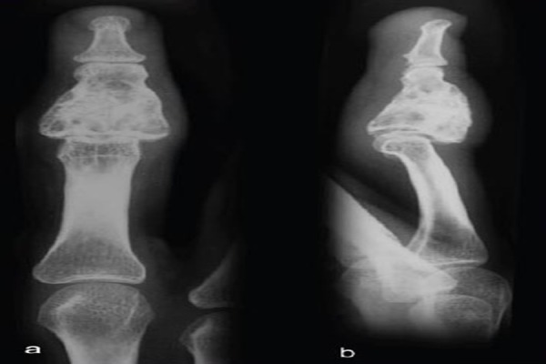
Case report: Authors report a case of epitheloid hemangioma of the middle phalanx in a young girl without any cutaneous manifestations. The lesion presented as a swollen middle finger, and plain radiographs showed a geographic area of destruction with cortical thinning and intra lesional calcifications. The case was managed by curettage and bone grafting. Histology confirmed this as a case of epitheloid hemangioma.
Conclusion: Epitheloid hemangioma should be considered in the differential diagnosis of hand masses with expansile lytic lesions with cortical thinning.
Keywords: Enchondroma; Epitheloid hemangioma; Hand; Vascular tumor.
Conflict of interest statement
Conflict of Interest: Nil
Figures
Similar articles
-
Case of Enchondroma of Left 4th Metacarpal of Hand Treated with Excision and Bone Grafting with Left 2nd Metatarsal of Foot: A Rare Case Report.J Orthop Case Rep. 2025 May;15(5):198-203. doi: 10.13107/jocr.2025.v15.i05.5610. J Orthop Case Rep. 2025. PMID: 40351631 Free PMC article.
-
Endoscopic Curettage and Bone Grafting of Enchondroma of Proximal Phalanx of Finger.Arthrosc Tech. 2023 Jul 10;12(8):e1335-e1340. doi: 10.1016/j.eats.2023.04.001. eCollection 2023 Aug. Arthrosc Tech. 2023. PMID: 37654891 Free PMC article.
-
Enchondroma protuberans of the hand: A case report.Radiol Case Rep. 2020 May 6;15(7):943-946. doi: 10.1016/j.radcr.2020.04.026. eCollection 2020 Jul. Radiol Case Rep. 2020. PMID: 32405332 Free PMC article.
-
Spinal epidural epitheloid hemangioma--case report and review of the literature.Pediatr Neurosurg. 2005 May-Jun;41(3):155-7. doi: 10.1159/000085875. Pediatr Neurosurg. 2005. PMID: 15995335 Review.
-
Histiocytoid hemangioma of bone: a benign lesion which may mimic angiosarcoma. Report of a case and review of literature.Skeletal Radiol. 1983;10(3):165-9. doi: 10.1007/BF00357772. Skeletal Radiol. 1983. PMID: 6356367 Review.
Cited by
-
Metastatic tumor of the hand of unknown primary origin.SAGE Open Med Case Rep. 2019 Mar 14;7:2050313X19836894. doi: 10.1177/2050313X19836894. eCollection 2019. SAGE Open Med Case Rep. 2019. PMID: 30899514 Free PMC article.
-
Angiolymphoid hyperplasia with eosinophilia involving the common digital artery of the hand: A case report and classification of upper limb lesions.Int J Surg Case Rep. 2017;39:84-87. doi: 10.1016/j.ijscr.2017.08.007. Epub 2017 Aug 10. Int J Surg Case Rep. 2017. PMID: 28822892 Free PMC article.
-
Thumb Distal Phalanx Giant Enchondroma. A Case Presentation and Literature Review.Maedica (Bucur). 2024 Mar;19(1):177-181. doi: 10.26574/maedica.2024.19.1.177. Maedica (Bucur). 2024. PMID: 38736932 Free PMC article.
References
-
- Moran CA, Suster S. Angiolymphoid hyperplasia with eosinophilia (Epitheloid hemangioma) of the lung. Am J Clin Pathol. 2005;123:762–765. - PubMed
-
- Cone RO, Hudkins P, Nguyen V, et al. Histiocytoid hemangioma of bone: A benign lesion which may mimic angiosarcoma- report of a case and review of literature. Skeletal Radiol. 1983;10:165–169. - PubMed
-
- Nielson GP, Srivastava A, Kattapuram S, et al. Epitheloid hemangioma of bone revisited. A study of 50 cases. Am J Surg Pathol. 2009;33(2):270–277. - PubMed
-
- Sung MK, Kim YS, Resnick D. Epitheloid hemangioma of bone. Skeletal Radiol. 2000;29:530–534. - PubMed
-
- Kleck CJ, Seidel MJ. Epitheloid hemangioma of the distal humerus with pathologic fracture. Orthopedics. 2012;35(1):e116–e119. - PubMed
Publication types
LinkOut - more resources
Full Text Sources