

Gossypiboma in Thigh- A Case Report
- PMID: 27298975
- PMCID: PMC4719318
- DOI: 10.13107/jocr.2250-0685.188
Gossypiboma in Thigh- A Case Report
Abstract
Introduction: The word Gossypiboma has been used for a retained surgical sponge/swab and is derived from gossypium(latin:cotton) and boma(Swahili-place of concealment). Other synonyms for this entity are textiloma, retained textile foreign body(RTFB)"/muslinoma. It is rare in muskulo-skeletal surgery.
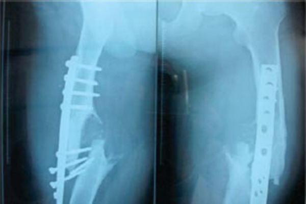
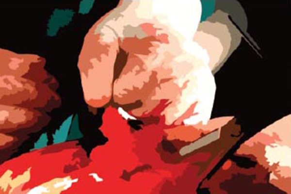
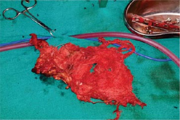
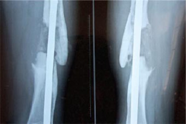
Case report: An eighteen year old boy was operated upon for failed plating of right femur. He had a globular swelling in mid thigh. There were no discharging sinuses, no signs/symptoms of infection. While operating on him to remove the failed implant and fix the fracture, while following standard procedures, we found a full size sponge embedded in the fracture site.
Conclusion: In all cases presenting with an incidental mass with/without sinus, Gossypiboma be kept in the differential diagnosis. Awareness of the condition is a must to diagnose such a rare condition. While operating one should make sure that no sponge is left inside-which can have serious medicolegal consequences.
Keywords: gossypiboma; osteoarticular surgery femur; retained sponges; textiloma.
Conflict of interest statement
Conflict of Interest: Nil
Figures
Similar articles
-
Oh, My Gauze !!!- A rare case report of laparoscopic removal of an incidentally discovered gossypiboma during laparoscopic cholecystectomy.Int J Surg Case Rep. 2020;72:643-646. doi: 10.1016/j.ijscr.2020.04.058. Epub 2020 May 16. Int J Surg Case Rep. 2020. PMID: 32513591 Free PMC article.
-
Gossypiboma: What Happens When a Mop is Left in a Thigh for ten Years.J Orthop Case Rep. 2020 Aug-Sep;10(5):12-15. doi: 10.13107/jocr.2020.v10.i05.1816. J Orthop Case Rep. 2020. PMID: 33312970 Free PMC article.
-
Intrathoracic gossypiboma presenting 47 years later as a purulent fistula: a case report.Surg Case Rep. 2022 Jun 24;8(1):123. doi: 10.1186/s40792-022-01479-6. Surg Case Rep. 2022. PMID: 35748964 Free PMC article.
-
[Gossypibomas in neurosurgery].Rev Neurol. 2019 Nov 1;69(9):377-382. doi: 10.33588/rn.6909.2019282. Rev Neurol. 2019. PMID: 31657450 Review. Spanish.
-
[Gossypiboma--retained textile foreign body].Chirurgia (Bucur). 2010 Nov-Dec;105(6):767-77. Chirurgia (Bucur). 2010. PMID: 21355175 Review. Romanian.
Cited by
-
Imaging Features of Soft Tissue Tumor Mimickers: A Pictorial Essay.Indian J Radiol Imaging. 2022 Aug 23;32(3):381-394. doi: 10.1055/s-0042-1756556. eCollection 2022 Sep. Indian J Radiol Imaging. 2022. PMID: 36177289 Free PMC article.
-
Gossypiboma and Total Hip Arthroplasty -- A Rare Accidental Finding Following a Periprosthetic Infection.J Orthop Case Rep. 2021 Jun;11(6):76-79. doi: 10.13107/jocr.2021.v11.i06.2266. J Orthop Case Rep. 2021. PMID: 35437497 Free PMC article.
-
Extremity gossypiboma mimicking sarcoma: case report and review.Skeletal Radiol. 2019 Apr;48(4):629-635. doi: 10.1007/s00256-018-3059-5. Epub 2018 Sep 10. Skeletal Radiol. 2019. PMID: 30203183
References
-
- Wilson C.P. Foreign bodies left in the abdomen after laparotomy. Gynecol. Tr. 1884;9:109–112.
-
- Rajagopal A, Martin J. Glossipiboma: “A surgeon's legacy”: Report of a case and review of literature. Dis Colon Rectum. 2002;45:119–20. PUBMED. - PubMed
-
- Mboti B, Gebhart M, Larsimont D, Abdelkafi K. Textiloma of thigh presenting as a sarcoma. Acta Orthop Belg. 2001;67:513–8. - PubMed
-
- Grieten M, Van Poppel H, Baert Al, Oyen R. Renal pseudo tumor due to a retained perirenal sponge: CT features. J Comput Assist Tomogr. 1992;16:305–7. PUBMED. - PubMed
-
- Kominani M, Fujikawa A, Tamura T, Naoi Y, Horikawa O. Retained surgical sponge in thigh: report of third known case in the limb. Radiat Med. 2003 Sep-Oct;21(5):220–2. PUBMED-Full text. - PubMed
Publication types
LinkOut - more resources
Full Text Sources