

Osteochondromas at Unusual Sites- Case Series with Review of Literature
- PMID: 27299127
- PMCID: PMC4845413
- DOI: 10.13107/jocr.2250-0685.376
Osteochondromas at Unusual Sites- Case Series with Review of Literature
Abstract
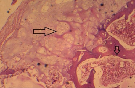
Introduction: Osteochondromas are benign tumours of the skeletal system. Their commonplace of occurrence is around growing ends of long bones like lower end of femur and upper end of tibia, but literature describing their incidence around flat bones of body like pelvis, scapula and small bones of hand, foot is rare.
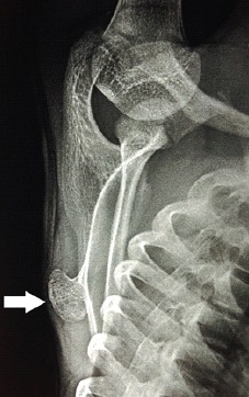
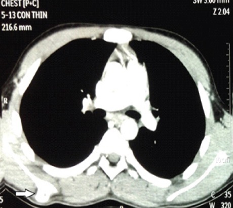
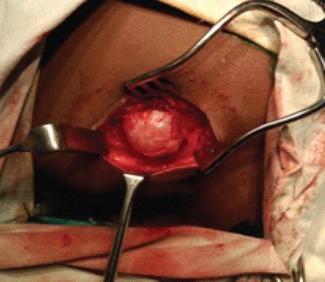
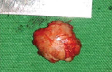
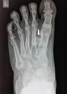
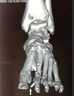
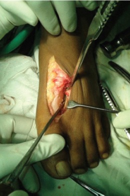
Case report: We describe two cases of osteochondromas at unusual sites, one on the dorsal aspect of scapula and other at the base of first metatarsal. Patient with scapular osteochondroma had difficulty in sleeping in supine position while that with metatarsal osteochondroma had discomfort while walking.
Conclusion: Depending on the site of occurrence, osteochondromas can give rise to different local symptoms. Possibility of osteochondroma should be kept in mind during differential diagnosis of bony swelling in flat bones as well as small bones.
Keywords: Unusual sites; bony swelling; osteochondroma.
Conflict of interest statement
Conflict of Interest: Nil
Figures
References
-
- Puri A, Agrawal M G. Current concepts in bone and soft tissue tumours. Textbook of orthopaedic oncology. Paras Medical Books Pvt. Ltd; pp. 72–77.
-
- Mahajan S, Mahajan N, Singh P, Shikari A, Sharma R. Scapula a rare localization of osteochondroma: a case report. The Internet Journal of Orthopedic Surgery. 2008;14(1)
-
- Essadki B, Moujtahid M, Lamine A, Fikry T, Essadki O, Zryouil B. Solitary osteochondroma of the limbs: clinical review of 76 cases and pathogenic hypothesis. ActaOrthopBelg. 2000;66:146–153. - PubMed
-
- Milgram JW. The origins of osteochondromas and enchondromas. A histopathologic study. Clin Orthop Relat Res. 1983;174:264–284. - PubMed
Publication types
LinkOut - more resources
Full Text Sources