Tuberculosis of Navicular Bone - A Rare Presentation
- PMID: 27299135
- PMCID: PMC4845421
- DOI: 10.13107/jocr.2250-0685.384
Tuberculosis of Navicular Bone - A Rare Presentation
Abstract
Introduction: Tuberculosis of Navicular bone is a rare entity. Osteoarticular tuberculosis of foot is uncommon and that of navicular bone is extremely rare. It is important to recognize skeletal tuberculosis in the initial stages as early treatment can effectively eliminate long-term morbidity.
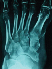
Case presentation: A 42 yrs old male presented to OPD with swelling and dull aching pain over dorsum of left foot. Radiograph of foot showed lytic puctate lesion in the navicular bone. Further investigations in the form of aspiration biopsy and ZN staining showed presence of multiple tuberculous bacilli. Anti-Kochs treatment was started immediately and patient was treated conservatively. Four drugs (HRZE) were given for a period of 12 months. Radiographs at 2 years follow-up showed a healed lesion.
Conclusion: TB navicular bone is a very rare condition and can be treated conservatively unless associated with metastatic changes or any other complications. Conservative treatment with AKT has excellent results without any complications.
Keywords: Navicular bone; Rare; Tuberculosis.
Conflict of interest statement
Conflict of Interest: Nil
Figures
References
-
- Ya JT, Yu CS. Diagnosis and Monitoring Treatment Response of Skeletal Tuberculosis of Foot by Three-phase Bone Scan: A Case Report. Ann Nucl Med Sci. 2010;23(3):175–180.
-
- Gupta R, Dhillon MS, Bahadur R, et al. Multifocal involvement of the foot in tuberculosis. Ind J Foot Surg. 2000;15:55–59.
-
- Mittal R, Gupta V, Rastogi S. Tuberculosis of the foot. J Bone Joint Surg(Br) 1999;81:997–1000. - PubMed
-
- Martni M. Adjrad Tuberculosis of the ankle and foot joint. In: Martini M, editor. Tuberculosis of the bones and joint. Berlin: Springer Verlag; 1988. pp. 143–149.
-
- Tuli SM. Tuberculosis of the skeletal system (bones, joints, spine and bursal sheaths) Second ed. New Delhi: Jaypee Brothers Medical Publishers (P) Ltd; 1991. pp. 3–122.
Publication types
LinkOut - more resources
Full Text Sources


