. 2016 Sep;34(9):2429-42.
doi: 10.1002/stem.2415. Epub 2016 Jul 4.

Cryopreserved Mesenchymal Stromal Cells Are Susceptible to T-Cell Mediated Apoptosis Which Is Partly Rescued by IFNγ Licensing

Affiliations

Cryopreserved Mesenchymal Stromal Cells Are Susceptible to T-Cell Mediated Apoptosis Which Is Partly Rescued by IFNγ Licensing

Raghavan Chinnadurai et al. Stem Cells. 2016 Sep.

Abstract

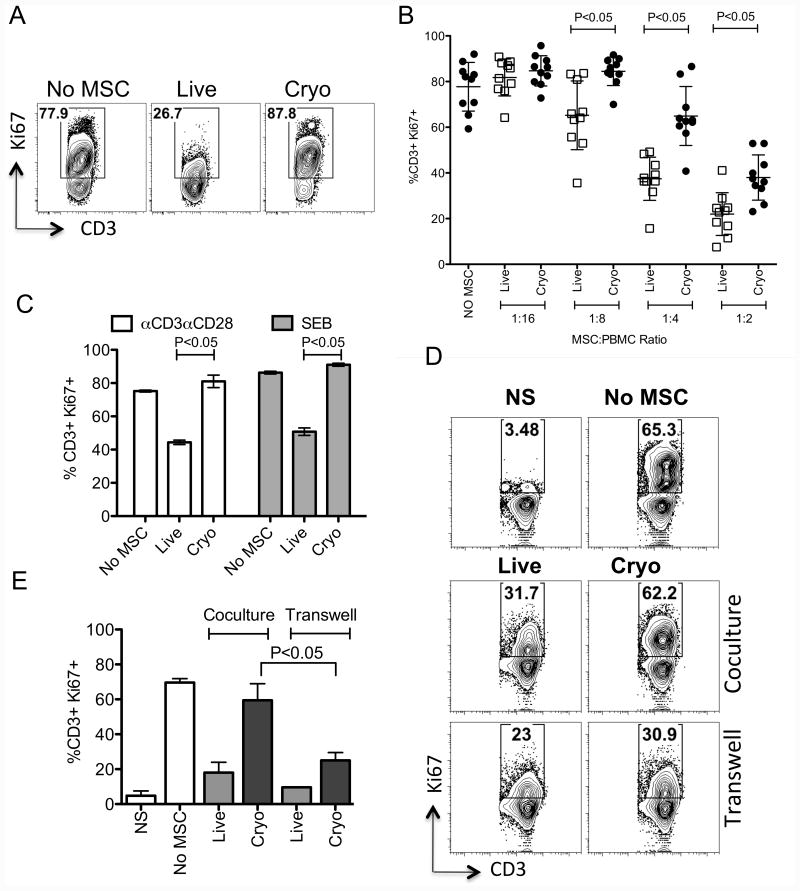
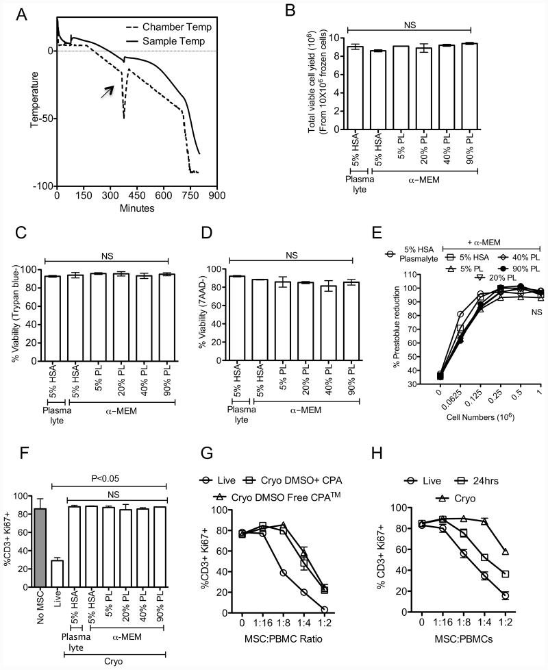
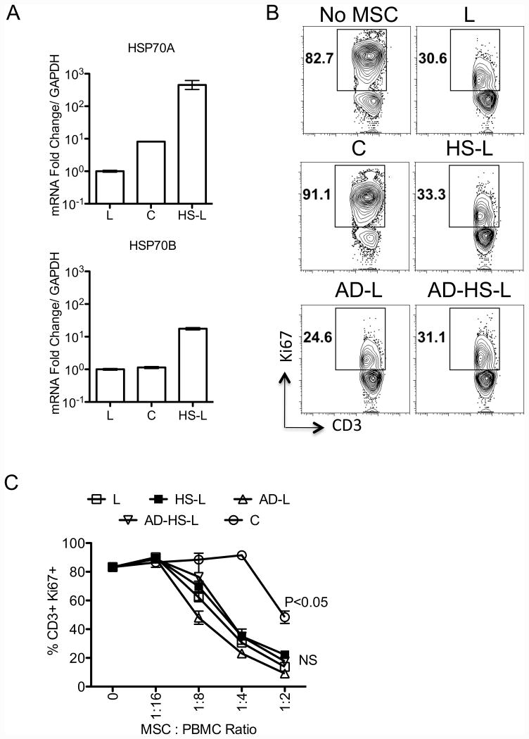
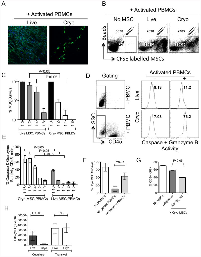
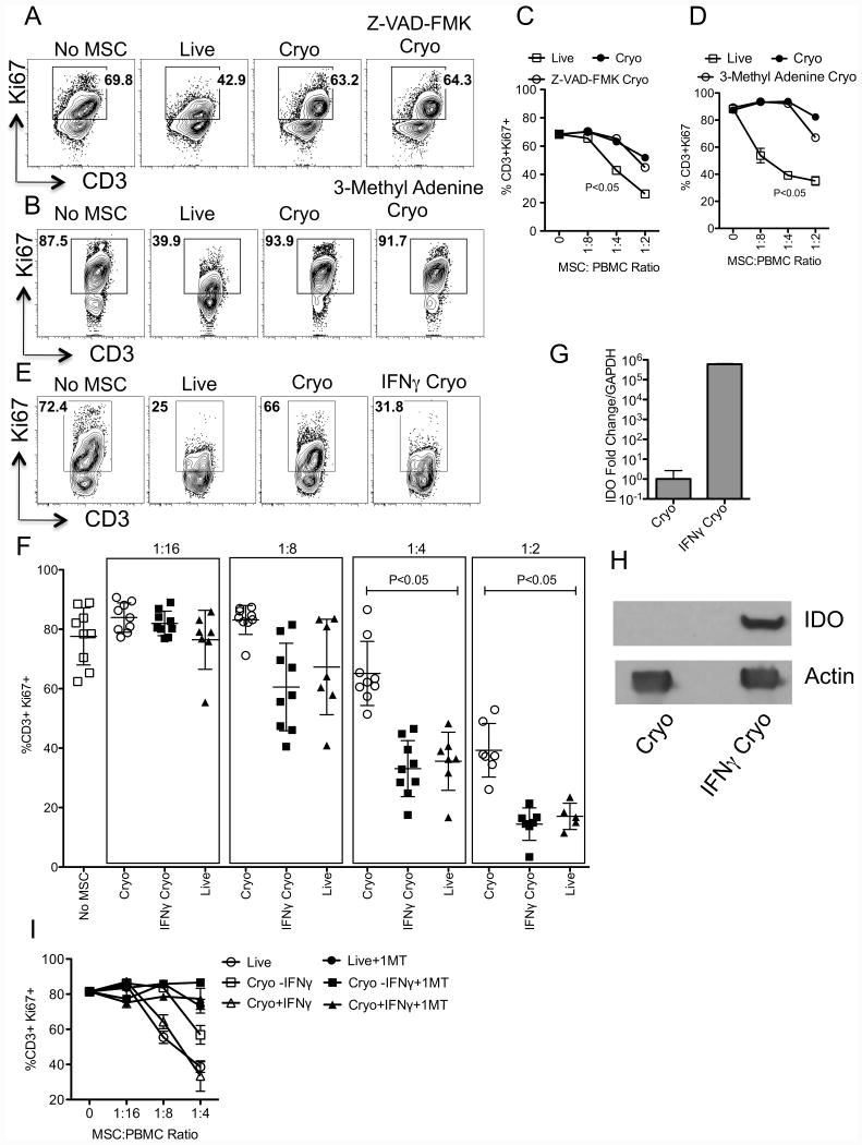
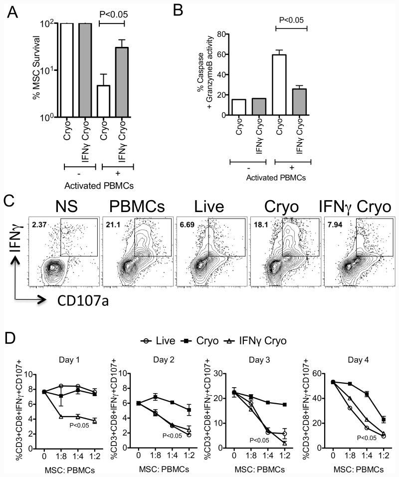
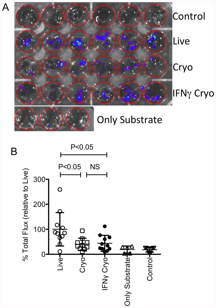
We have previously demonstrated that cryopreservation and thawing lead to altered Mesenchymal stromal cells (MSC) functionalities. Here, we further analyzed MSC's fitness post freeze-thaw. We have observed that thawed MSC can suppress T-cell proliferation when separated from them by transwell membrane and the effect is lost in a MSC:T-cell coculture system. Unlike actively growing MSCs, thawed MSCs were lysed upon coculture with activated autologous Peripheral Blood Mononuclear Cells (PBMCs) and the lysing effect was further enhanced with allogeneic PBMCs. The use of DMSO-free cryoprotectants or substitution of Human Serum Albumin (HSA) with human platelet lysate in freezing media and use of autophagy or caspase inhibitors did not prevent thaw defects. We tested the hypothesis that IFNγ prelicensing before cryobanking can enhance MSC fitness post thaw. Post thawing, IFNγ licensed MSCs inhibit T cell proliferation as well as fresh MSCs and this effect can be blocked by 1-methyl Tryptophan, an Indoleamine 2,3-dioxygenase (IDO) inhibitor. In addition, IFNγ prelicensed thawed MSCs inhibit the degranulation of cytotoxic T cells while IFNγ unlicensed thawed MSCs failed to do so. However, IFNγ prelicensed thawed MSCs do not deploy lung tropism in vivo following intravenous injection as well as fresh MSCs suggesting that IFNγ prelicensing does not fully rescue thaw-induced lung homing defect. We identified reversible and irreversible cryoinjury mechanisms that result in susceptibility to host T-cell cytolysis and affect MSC's cell survival and tissue distribution. The susceptibility of MSC to negative effects of cryopreservation and the potential to mitigate the effects with IFNγ prelicensing may inform strategies to enhance the therapeutic efficacy of MSC in clinical use. Stem Cells 2016;34:2429-2442.

Keywords: Autophagy; Cryopreservation; DMSO; Heat shock; Immune suppression; Indoleamine 2,3-dioxygenase; Mesenchymal stromal cells; T cell responses; Thawing; actin.


Figures

Figure 1
Figure 1. Frozen-thawed human MSCs display cell contact-dependent attenuated immunosuppressive properties on T cells
PBMCs co-cultured in the presence and absence of MSCs derived from actively growing culture (Live) or thawed from cryopreservation (Cryo) were stimulated with SEB. 4 days post, T cell proliferation was measured by Ki67 intracellular staining. (A) Representative FACS plot and (B) cumulative % of T cell proliferation (CD3+Ki67+) in the presence of variable MSC and PBMC ratio is shown. Cumulative is plotted from multiple independent experiments tested with unique PBMC and MSC donors. (C) Live and Cryo MSCs were cocultured with SEB and aCD3aCD28 activated PBMCs. 4 days post culture, T cell proliferation was evaluated. Live and cryo MSCs were cultured with SEB activated T cells in a contact and non-contact dependent coculture and transwell system respectively. (A) Representative FACS plot and (B) cumulative % of T cell proliferation (CD3+Ki67+) in the coculture and transwell system is shown. Cumulative is plotted from three independent experiments with unique MSC and PBMC donors. P value <0.05 was considered statistically significant based upon two-tail T-tests.
Figure 2
Figure 2. Freeze-thawing attenuates immunosuppressive properties of human MSCs independent of freezing methods
(A) Freezing program that was used to cryopreserve MSCs in a step down freezer. Chamber and sample temperature during step-down freezing is shown. An arrow shows alignment of “heat of fusion” between chamber and sample temperature. Live or cryopreserved MSCs with the indicated formulations of cryoprotectant were thawed (B) Total viable cell yield (C) Trypan blue viability (D) 7-AAD negative viability, (E) Prestoblue reduction potential was investigated. Cryo MSC populations were cocultured with SEB activated PBMCs and Live MSCs were used as controls. 4 days post culture (F) T cell proliferation (%CD3+Ki67+) was measured in flow cytometry. (G) Live or Cryo MSCs cryopreserved with 10% DMSO or DMSO free cryoprotectant, (H) Live, thawed and 24-hour culture rescued MSCs were and subjected to test their inhibitory effect on T cell proliferation as indicated above. T cell proliferation was measured 4 days post culture. Representative experiments are shown from two independent experiments performed on two unique MSC donors with independent methods. One-way ANOVA using the Tukey's Multiple Comparison Test was used to compare multiple groups. P value <0.05 was considered statistically significant.
Figure 3
Figure 3. Freeze-thawing induced heat shock response and disruption of actin polymerization do not modulate human MSC's immunosuppressive properties
Live (L) MSCs were subjected in to three treatments namely, Cytochalasin D (2mM) for 2 hours to depolymerize actin cytoskeleton (AD-L), cultured in a 42C incubator for three hours to induce heat shock response (HS-L) and both (AD-HS-L). (A) Cryo (C), L, and HS-L MSCs were tested for the mRNA expression of heat shock proteins HSP70A, HSP70B. Expression level of HSP70A, HSP70B mRNA relative to GAPDH was evaluated by the quantitative SYBR green real time PCR. Delta-delta CT method was applied to calculate the fold induction of HSP70A, HSP70B over the Live (L) control. MSCs derived from conditions L, C, HS-L, AD-L, AD-HS-L were cocultured with SEB activated PBMCs with the indicated ratios for 4days and T cell proliferation was measured by Ki67 intracellular staining. (B) Representative FACS plot and (C) Dose dependent effect of MSCs on of T cell proliferation (CD3+Ki67+) is shown. A representative experiment is shown from two independent experiments performed on two unique MSC donors. P value <0.05 was considered statistically significant based upon two-tail T-tests.
Figure 4
Figure 4. Frozen-thawed MSCs are susceptible to lysis by activated T cells
CFSE labeled Live and Cryo MSCs were cocultured with SEB activated PBMCs in indicated ratios. (A) Microscopic images show the survival of CFSE labeled (Green) Live and Cryo MSCs in the coculture. Nuclei were stained with DAPI (blue). Plate bound MSCs from the coculture were trypsinized and event counts were recorded in flow cytometry with the normalization of counting beads. Live and Cryo MSC count in the absence of PBMCs were used for normalization and calculation of % survival of Live and Cryo MSCs in the presence of PBMCs. (B) Representative FACS plot and (C) dose dependent effect of activated PBMCs on Live and Cryo MSC's survival is shown. Serine protease activity was measured on Live and Cryo MSCs cocultured with/without activated PBMCs. (D) Representative FACS plot and (E) dose dependent effect of activated PBMCs on the serine protease activity in Live and Cryo MSCs were shown. (F) % Survival of CFSE labeled thawed MSCs cocultured with autologous and allogeneic PBMCs were shown. (G) Effect of Cryo MSCs in inhibiting autologous and allogeneic T cell proliferation (CD3+Ki67+) is shown. (H) Relative CFSE count of Live and Cryo MSCs in the presence of activated PBMCs cocultured or separated by a transwell system is shown. A representative experiment is shown from at least three independent experiments performed on one to three unique MSC donors. P value <0.05 was considered statistically significant based upon two-tail T-tests.
Figure 5
Figure 5. IFNγ prelicensing but not inhibitors of autophagy and caspase rescues frozen-thawed MSC's defective immunosuppressive properties
+/- IFNγ, Z-VAD-FMK, 3-Methyl adenine(3-MA) pretreated MSCs were cryopreserved and thawed to compare with live MSC's immunosuppressive potential. MSC populations (Live, Cryo and IFNγ/Z-VAD-FMK/3-MA Cryo) were cocultured with SEB activated PBMCs in indicated ratios. PBMC and MSC numbers were kept constant and variable for escalating ratios. 4 days post, T cell proliferation was measured by flow cytometry. Representative and dose dependent effect of (A, C) Z-VAD-FMK and (B, D) 3-MA treated thawed MSCs on T cell proliferation (CD3+Ki67+) is shown. Similar results were obtained in another experiment. (E) Representative FACS plot and (F) cumulative % of T cell proliferation (CD3+Ki67+) with +/-IFNγ Cryo, Live MSC and PBMC ratio is shown. Cumulative is plotted from multiple independent experiments tested with unique PBMC and MSC donors. (G) +/- IFNγ licensed cryopreserved human MSCs were thawed and RNA was extracted to quantitate the expression levels of IDO mRNA by quantitative sybr-green real time PCR. GAPDH mRNA levels were used as an internal control. Delta-delta CT method was applied to calculate the fold change. (H) Western blot analysis to show the IDO expression at protein levels in IFNγ cryo MSCs. Actin was used as an internal control. (I) Live, cryo and IFNγ cryo MSCs were cocutured with activated PBMCs in the presence and absence of IDO blocker, 1-Methyl Tryptophan (1MT). 4 days post, T cell proliferation was measured by flow cytometry. P value <0.05 was considered statistically significant based upon two-tail T-tests.
Figure 6
Figure 6. IFNγ prelicensing rescues frozen-thawed MSC's survival through inhibition of cytotoxic T cell degranulation
CFSE labeled +/- IFNγ Cryo MSCs were cocultured with SEB activated PBMCs for 4 days in seeding ratio of 1:4. Trypsinzied CFSE+ cells were counted in flow cytometry using counting bead normalization. +/- IFNγ Cryo MSC count in the absence of PBMCs were used for normalization and calculation of % survival of +/- IFNγ Cryo MSCs in the presence of PBMCs. Live MSC count in the presence of PBMCs were used for normalization and calculation of % survival of +/- IFNγ Cryo MSCs with PBMCs. (A) Relative % survival of live, cryo and IFNγ cryo MSCs cocultured with SEB activated PBMCs is shown. (B) Percentage serine protease activity in cryo and IFNγ cryo MSCs cocultured with activated SEB cells is shown. SEB activated PBMCs were cocultured in the presence and absence of live, cryo and IFNγ cryo MSCs. 12-14 hours prior to the indicated time point BFA, monensin and antibody to CD107 were added to the culture. Cells were subsequently stained with antibodies to CD3, CD8 and IFNγ for flow cytometry. (C) Representative and (D) kinetics of % of CD3+CD8+CD107+IFNγ+T cells is shown. Similar results were obtained in a repeat experiment with another MSC donor. P value <0.05 was considered statistically significant based upon two-tail T-tests.
Figure 7
Figure 7. IFNγ prelicensing does not rescue frozen-thawed MSC's defective engraftment properties
1×10⁶ of cryo, IFNγ cryo and Live MSCs derived from luciferase transgenic B6 animals were injected intravenously into C57BL/B6 mice via the tail vein. 24 hours post infusion, the animals were sacrificed and the lungs were excised placed in 24 well plate. D-Luciferin substrate was added at the same time to each well and the plate was subjected to bioluminescence imaging. (A) Representative imaging and (B) cumulative relative % binding of MSCs are shown from two independent experiments (n=5 or 6 animals/group). Cumulative Mean±SD are shown with a P value of <0.05 was considered statistically significant in prism software. One outlier in cryo group was removed based on the extreme studentized deviate method or Grubbs' test in graph pad prism software.

References

    1. Galipeau J, Krampera M. The challenge of defining mesenchymal stromal cell potency assays and their potential use as release criteria. Cytotherapy. 2015;17:125–127. - PubMed
    1. Stagg J, Galipeau J. Mechanisms of immune modulation by mesenchymal stromal cells and clinical translation. Curr Mol Med. 2013;13:856–867. - PubMed
    1. Le Blanc K, Mougiakakos D. Multipotent mesenchymal stromal cells and the innate immune system. Nat Rev Immunol. 2012;12:383–396. - PubMed
    1. Pittenger MF, Mackay AM, Beck SC, et al. Multilineage potential of adult human mesenchymal stem cells. Science. 1999;284:143–147. - PubMed
    1. Dominici M, Le Blanc K, Mueller I, et al. Minimal criteria for defining multipotent mesenchymal stromal cells. The International Society for Cellular Therapy position statement. Cytotherapy. 2006;8:315–317. - PubMed

MeSH terms