. 2016 Jun 14;11(6):e0157390.
doi: 10.1371/journal.pone.0157390. eCollection 2016.

Malaria Parasite CLAG3, a Protein Linked to Nutrient Channels, Participates in High Molecular Weight Membrane-Associated Complexes in the Infected Erythrocyte

Affiliations

Malaria Parasite CLAG3, a Protein Linked to Nutrient Channels, Participates in High Molecular Weight Membrane-Associated Complexes in the Infected Erythrocyte

Kayvan Zainabadi. PLoS One. .

Abstract

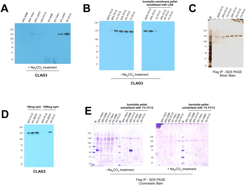
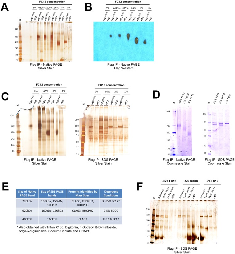
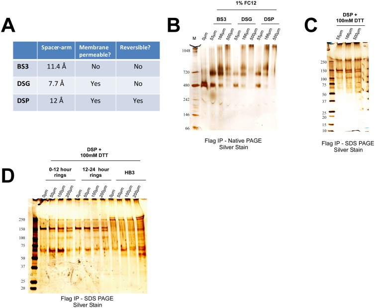
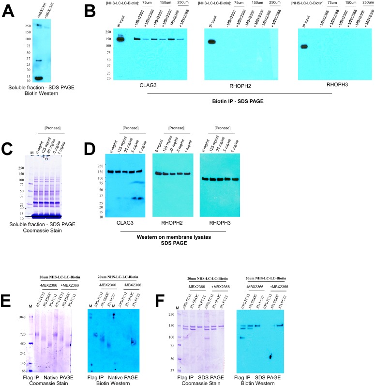
Malaria infected erythrocytes show increased permeability to a number of solutes important for parasite growth as mediated by the Plasmodial Surface Anion Channel (PSAC). The P. falciparum clag3 genes have recently been identified as key determinants of PSAC, though exactly how they contribute to channel function and whether additional host/parasite proteins are required remain unknown. To begin to answer these questions, I have taken a biochemical approach. Here I have used an epitope-tagged CLAG3 parasite to perform co-immunoprecipitation experiments using membrane fractions of infected erythrocytes. Native PAGE and mass spectrometry studies reveal that CLAG3 participate in at least three different high molecular weight complexes: a ~720kDa complex consisting of CLAG3, RHOPH2 and RHOPH3; a ~620kDa complex consisting of CLAG3 and RHOPH2; and a ~480kDa complex composed solely of CLAG3. Importantly, these complexes can be found throughout the parasite lifecycle but are absent in untransfected controls. Extracellular biotin labeling and protease susceptibility studies localize the 480kDa complex to the erythrocyte membrane. This complex, likely composed of a homo-oligomer of 160kDa CLAG3, may represent a functional subunit, possibly the pore, of PSAC.


Conflict of interest statement

Competing Interests: The author has declared that no competing interests exist.

Figures

Fig 1
Fig 1. Solubilization and immunoprecipitation of membrane-associated CLAG3.
(A) Western blot analysis shows that the zwitterionic detergents, lauryldimethyl amine oxide (LDAO) and fos-choline 12 (FC12), are able to solubilize integral CLAG3 at 0.5% (w/v) from sodium carbonate pH 11 (Na2CO3) treated trophozoite-infected erythrocyte membranes. (B) FC12 solubilizes total CLAG3 from (non-carbonate treated) membranes at concentrations 0.1% or above, leaving almost nothing behind in the insoluble membrane pellet. (LDS: lithium dodecyl sulfate). (C) Immunoprecipitation (IP) of CLAG3 from a previously described FLAG-tagged CLAG3 parasite (HB33REC) results in the appearance of the expected 160kDa CLAG3 band at FC12 concentrations ≥ 0.1%, and two additional faint bands (150kDa and 100kDa) at FC12 concentrations ≤ 0.05% [8]. (D) Substantially more CLAG3 remains in the detergent-soluble supernatant following a 16,000xg spin to pellet insoluble material as compared to a traditional 100,000xg spin for all detergents tested (SDOC: sodium deoxycholate). (E) IP experiments were repeated incorporating the slower 16,000xg spin. All detergents tested (with the exception of 0.5% FC12) give rise to the aforementioned three bands in non-carbonate (-Na2CO3) treated membranes, but fail to do so in membranes treated with sodium carbonate (+Na2CO3). As evidenced by the right half of each gel, the only detergent able to fully solubilize CLAG3 from membranes (and leave nothing behind in the insoluble pellet) is 0.5% FC12. The prominent 60kDa band corresponds to heavy chain IgG.
Fig 2
Fig 2. CLAG3 forms high molecular weight membrane-associated complexes.
(A) Blue Native PAGE analysis of FLAG IP eluates obtained from a FLAG-tagged CLAG3 parasite (HB33REC) and an isogenic untagged HB3 negative control [8]. Two distinct bands can be seen in HB33REC that are absent in HB3: a ~720kDa band for concentrations of FC12 ≤ 0.05% and a ~480kDa band for concentrations of FC12 ≥ 0.1%. (B) FLAG Western blot confirms that the 720kDa and 480kDa bands contain CLAG3. (C) SDS PAGE analysis shows that upon addition of SDS and 2-mercaptoethanol the 720kDa complex dissociates to three proteins (CLAG3, RHOPH2, and RHOPH3 as determined by mass spectrometry), while the 480kDa complex dissociates to only one protein (CLAG3). (D) Sodium deoxycholate (SDOC) at 0.5% (w/v) yields an intermediate ~620kDa Native PAGE band that contains CLAG3 and RHOPH2, but not RHOPH3 (as determined by SDS PAGE and mass spectrometry). (E) A summary of the mass spectrometry results for the indicated Native and SDS PAGE bands. (F) IP experiments on tightly synchronized parasites show that CLAG3, RHOPH2 and RHOPH3 appear to associate faithfully in early (0–6 hours post-invasion), mid-stage (12–16 hours post-invasion), or late rings/early trophozoites (24–30 hours post-invasion). The prominent 30kDa band corresponds to light chain IgG.
Fig 3
Fig 3. Cross-linking studies with NHS-esters confirm the existence of CLAG3 complexes.
(A) The properties of the three different N-hydroxysulfosuccinimide esters (NHS-esters) used in this study: Bis(sulfosuccinimidyl) suberate (BS3), Disuccinimidyl glutarate (DSG), and Dithiobis (succinimidyl propionate) (DSP). (B) The 480kDa complex is observed with the use of 1% FC12, whereas addition of the three different NHS-esters results in the appearance of higher molecular weight complexes in a dose dependent manner (as determined by Native PAGE). (C) DSP cross-link was reversed with the addition of 100mM Dithiothreitol (DTT) and bands separated on SDS PAGE. The three prominent bands observed are consistent in size with CLAG3, RHOPH2, and RHOPH3. The band near 250kDa was subsequently found to also be present in HB3 negative control parasites (see next panel). (D) IP experiments on tightly synchronized parasites obtained without enrichment and cross-linked with DSP demonstrate no appreciable differences in the association CLAG3, RHOPH2, and RHOPH3 in early (0–12 hour) or late (12–24 hour) rings (when PSAC is inactive and active, respectively). HB3 was used as a negative control.
Fig 4
Fig 4. The 480kDa complex composed of CLAG3 localizes to the erythrocyte membrane.
(A) Incubation of infected erythrocytes with extracellular NHS-LC-LC-Biotin results in labeling of intracellular proteins due to traversal through PSAC, which is prevented in the presence of PSAC inhibitor, MBX2366. (B) Membranes of biotin-labeled cells were solubilized with 1% FC12 and subjected to biotin IP, followed by Western blot for the indicated proteins. As indicated by the IP inputs, 1% FC12 is able to equally solubilize CLAG3, RHOPH2 and RHOPH3. However, only CLAG3 is found in the biotin IP eluate indicating localization to the erythrocyte surface. (C) Treatment with extracellular pronase-E does not result in the cleavage of intracellular proteins. (D) Extracellular pronase-E results in the appearance of a cleavage fragment for CLAG3, but not RHOPH2 and RHOPH3, in a dose-dependent fashion. C-terminal antibodies were used for all three proteins. (E) Extracellular biotin labeling results in labeling of the 480kDa complex in the presence of MBX2366 indicating that this complex localizes to the erythrocyte membrane. (F) Consistent with the above results, Western blot analysis with biotin antibody shows that only the CLAG3 obtained with 0.5% FC12 (corresponding to the 480kDa complex) labels with biotin in the presence of MBX2366.

References

    1. Homewood CA, Neame KD. Malaria and the permeability of the host erythrocyte. Nature. 1974;252: 718–719 - PubMed
    1. Ginsburg H, Kutner S, Zangwil M, Cabantchik ZI. Selectivity properties of pores induced in host erythrocyte membrane by Plasmodium falciparum. Effect of parasite maturation. Biochim Biophys Acta. 1986;861:194–196. - PubMed
    1. Staines HM, Rae C, Kirk K. Increased permeability of the malaria-infected erythrocyte to organic cations. Biochim. Biophys. Acta. 2000;1463:88–98 - PubMed
    1. Ginsburg H, Kutner S, Krugliak M, Cabantchik ZI. Characterization of permeation pathways appearing in the host membrane of Plasmodium falciparum infected red blood cells. Mol. Biochem. Parasitol. 1985;14:313–322 - PubMed
    1. Desai SA, Bezrukov SM, Zimmerberg J. A voltage-dependent channel involved in nutrient uptake by red blood cells infected with the malaria parasite. Nature. 2000;406:1001–1005 - PubMed

MeSH terms

Substances

LinkOut - more resources