. 2016 Jun 14;11(6):e0157537.
doi: 10.1371/journal.pone.0157537. eCollection 2016.

A Farnesyltransferase Acts to Inhibit Ectopic Neurite Formation in C. elegans

Affiliations

A Farnesyltransferase Acts to Inhibit Ectopic Neurite Formation in C. elegans

David Carr et al. PLoS One. .

Abstract

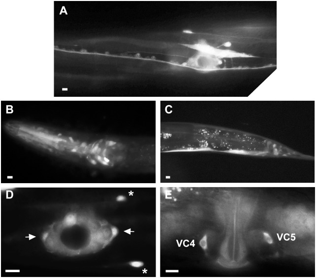
Genetic pathways that regulate nascent neurite formation play a critical role in neuronal morphogenesis. The core planar cell polarity components VANG-1/Van Gogh and PRKL-1/Prickle are involved in blocking inappropriate neurite formation in a subset of motor neurons in C. elegans. A genetic screen for mutants that display supernumerary neurites was performed to identify additional factors involved in this process. This screen identified mutations in fntb-1, the β subunit of farnesyltransferase. We show that fntb-1 is expressed in neurons and acts cell-autonomously to regulate neurite formation. Prickle proteins are known to be post-translationally modified by farnesylation at their C-terminal CAAX motifs. We show that PRKL-1 can be recruited to the plasma membrane in both a CAAX-dependent and CAAX-independent manner but that PRKL-1 can only inhibit neurite formation in a CAAX-dependent manner.


Conflict of interest statement

Competing Interests: The authors have declared that no competing interests exist.

Figures

Fig 1
Fig 1. A genetic screen for VC4 and VC5 neurite outgrowth defective (Nde) mutants.
(A) Worm schematic showing the position of VC1–6 (ventral view). VC4 and VC5 neurons extend neurites along a LR axis and VC1–3 and VC6 extend neurites along the AP body axis. (A) Wild-type VC4 and VC5. (B) In prkl-1 and vang-1 (not shown) mutants, VC4 and VC5 display ectopic neurites (arrows) directed along the AP axis. (D) Schematic outline of a forward genetic screen for VC4 and VC5 neurite outgrowth defective (nde) mutants. (E) Quantification of ectopic neurite defects in nde mutants. Graph shows the percentage of VC4 and VC5 neurons (pooled) displaying at least one ectopic neurite. Error bars represent the 95% confidence interval of the proportion. For each genotype, n>160. (F and G) Representative images of VC4 and VC5 in nde-4 (F) and nde-5 (G) mutants. Ectopic neurites are marked with arrows. All lines contain the cyIs4[Pcat-1::GFP] reporter background. All images are ventral views. Scale bars, 10μm.
Fig 2
Fig 2. The nde-4 gene encodes the farnesyltransferase (FTase) β subunit (fntb-1).
(A) nde-4 mutants were mapped to a small interval on chromosome V and rescued with a genomic fragment containing the fntb-1 (F23B12.6) gene and an FNTB-1::GFP-translational fusion driven from the fntb-1 promoter. Promoter and genomic regions used in rescuing constructs and a Pfntb-1::GFP transcriptional reporter are delineated. (B) Quantification of VC4 and VC5 ectopic neurite rescue in nde-4(zy7) mutants by fntb-1 genomic, FNTB-1::GFP and unc-4 promoter driven FNTB-1 constructs. Representative extrachromosomal lines are shown. Error bars represent the standard error of the proportion, n>200. *p<0.001, t test.
Fig 3
Fig 3. FNTB-1 missense mutations mapped to human FTase.
(A) Crystal structure of the human protein FTase (PDB ID: 1S63) heterodimer (α subunit; red and β subunit; blue). Yellow circles mark the location of residues C104, G250, S301 and Q336 of the worm FTase. Lipid substrate farnesyl diphosphate, (FPP: isoprenoid moiety, green; diphosphate, red) and zinc ion (grey sphere) mark the site of catalysis. (B) Enlarged view of the FTase active site. G250 (yellow surface representation) makes van der Waals contact with FPP (grey surface). Any mutation at position 250 would result in a larger side chain that would affect FPP binding. (C) Enlarged view around residue C92 in worm FTase (C104 human FTase; green stick). The four major rotamers for the C104Y mutation and their corresponding van der Waals surfaces are shown in yellow. A van der Waals surface calculated around the alpha subunit (red) and beta subunit (blue) is shown. Any tyrosine rotamer results in a steric class with the protein. This mutation is predicted to compromise the packing and the stability of the enzyme.
Fig 4
Fig 4. fntb-1 is expressed in VC4 and VC5 neurons.
(A) Pfntb-1::GFP expression in the mid-body region of an L4 hermaphrodite. Pfntb-1::GFP expression in the head (B) and tail (C) region of an L4 hermaphrodite. The fntb-1 promoter is active in neurons, vulval and rectal epithelial cells and in muscle cells. (D) Pfntb-1::GFP transcriptional activity in VC4 and VC5 (arrows), HSNs (asterisks) and vulval epithelial cells at the mid-L4 stage vulval region. (E) Adult vulval region showing FNTB-1::GFP protein fusion predominantly localized to the cytoplasm of VC4 and VC5 neurons. Panels A, D and E show ventral views. Panels B and C show side views. Scale bars, 10μm.
Fig 5
Fig 5. The PRKL-1 CAAX motif is necessary for normal VC4 and VC5 morphology.
(A) Schematic of PRKL-1 showing conserved domains (black boxes) and C-terminal CAAX motif. (B) Expression of CAAX deleted PRKL-1 does not rescue VC4 and VC5 morphology defects in prkl-1 mutants as well as full length PRKL-1. Replacement of the PRKL-1 CAAX motif with the CNIM motif from MIG-2 resembles wildtype PRKL-1 in rescuing VC4 and VC5 morphology defects. (C) PRKL-1 overexpression can rescue VC morphology defects in vang-1 mutants but not fntb-1 mutants. All transgenic lines in B and C contain unc-4 promoter-driven PRKL-1 constructsand all lines contain the cyIs4[Pcat-1::GFP] reporter background. Error bars represent the 95% confidence interval of the proportion, n>150. *p<0.001, t test.
Fig 6
Fig 6. The PRKL-1 CAAX motif is important for membrane localization.
(A) Transgene expression of a GFP::PRKL-1 fusion shows punctate localization on the plasma membrane of VC4 and VC5 (many puncta) at the early L4 stage. Representative images showing the localization of a CAAX-deleted PRKL-1::GFP fusion in a wild-type (wt) background (B) and full length GFP::PRKL-1 in a vang-1 mutant background (C). In both panels B and C, a localization pattern resembling full length PRKL-1 (many puncta) or diminished membrane localization (few or no puncta, arrows) are observed. (D) Expression of a CAAX-deleted PRKL-1 construct in a vang-1 mutant background shows loss of plasma membrane localization in VC4 and VC5 (few/no puncta). (E) Quantification of full length GFP::PRKL-1 and GFP::PRKL-1 ΔCAAX membrane distribution in wt and vang-1 mutants in early L4 stage VC4 and VC5.

Similar articles

Cited by

References

    1. van Beuningen SFB, Will L, Harterink M, Chazeau A, van Battum EY, Frias CP, et al. TRIM46 Controls Neuronal Polarity and Axon Specification by Driving the Formation of Parallel Microtubule Arrays. Neuron. 2015; 1208–1226. 10.1016/j.neuron.2015.11.012 - DOI - PubMed
    1. Bradke F, Dotti CG. The role of local actin instability in axon formation. Science. 1999;283: 1931–4. Available: http://www.ncbi.nlm.nih.gov/pubmed/10082468 - PubMed
    1. Flynn KC, Hellal F, Neukirchen D, Jacob S, Tahirovic S, Dupraz S, et al. ADF/cofilin-mediated actin retrograde flow directs neurite formation in the developing brain. Neuron. 2012;76: 1091–107. 10.1016/j.neuron.2012.09.038 - DOI - PubMed
    1. Kwiatkowski AV, Rubinson DA, Dent EW, Edward van Veen J, Leslie JD, Zhang J, et al. Ena/VASP Is Required for Neuritogenesis in the Developing Cortex. Neuron. 2007;56: 441–455. 10.1016/j.neuron.2007.09.008 - DOI - PubMed
    1. Adler CE, Fetter RD, Bargmann CI. UNC-6/Netrin induces neuronal asymmetry and defines the site of axon formation. Nat Neurosci. 2006;9: 511–8. 10.1038/nn1666 - DOI - PMC - PubMed

MeSH terms

Substances

LinkOut - more resources