. 2016 Jun 14;12(6):e1005660.
doi: 10.1371/journal.ppat.1005660. eCollection 2016 Jun.

Immunosuppressive Yersinia Effector YopM Binds DEAD Box Helicase DDX3 to Control Ribosomal S6 Kinase in the Nucleus of Host Cells

Affiliations

Immunosuppressive Yersinia Effector YopM Binds DEAD Box Helicase DDX3 to Control Ribosomal S6 Kinase in the Nucleus of Host Cells

Laura Berneking et al. PLoS Pathog. .

Abstract

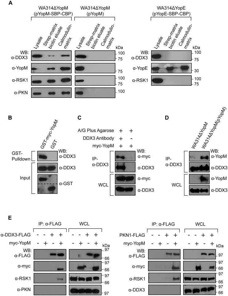
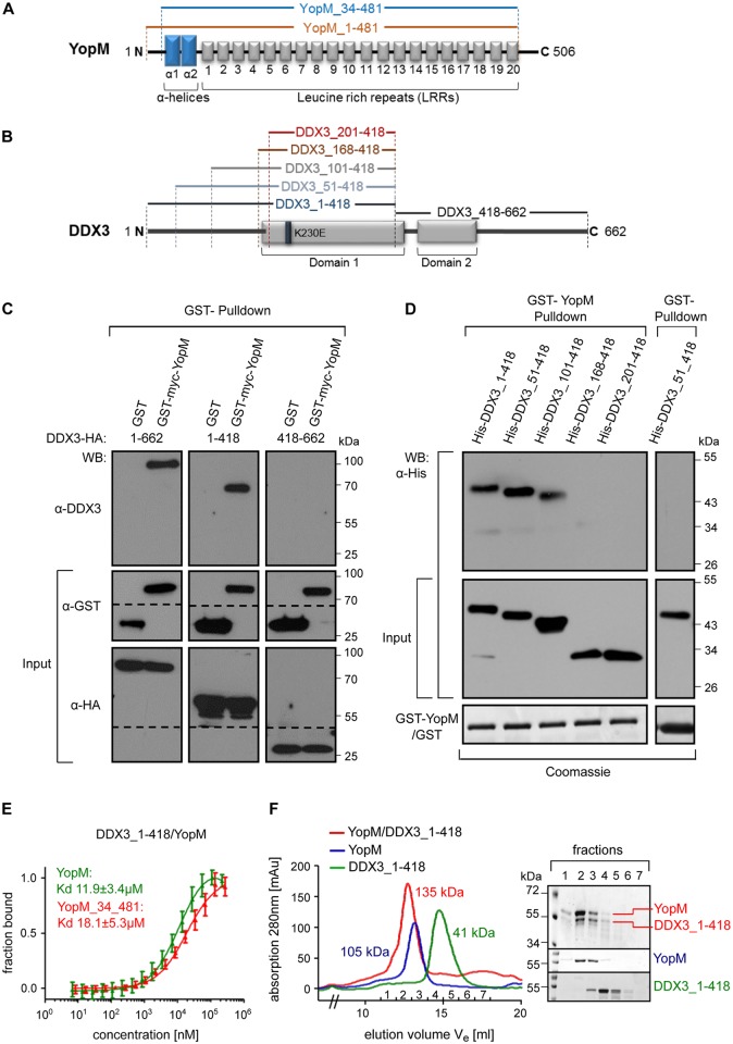
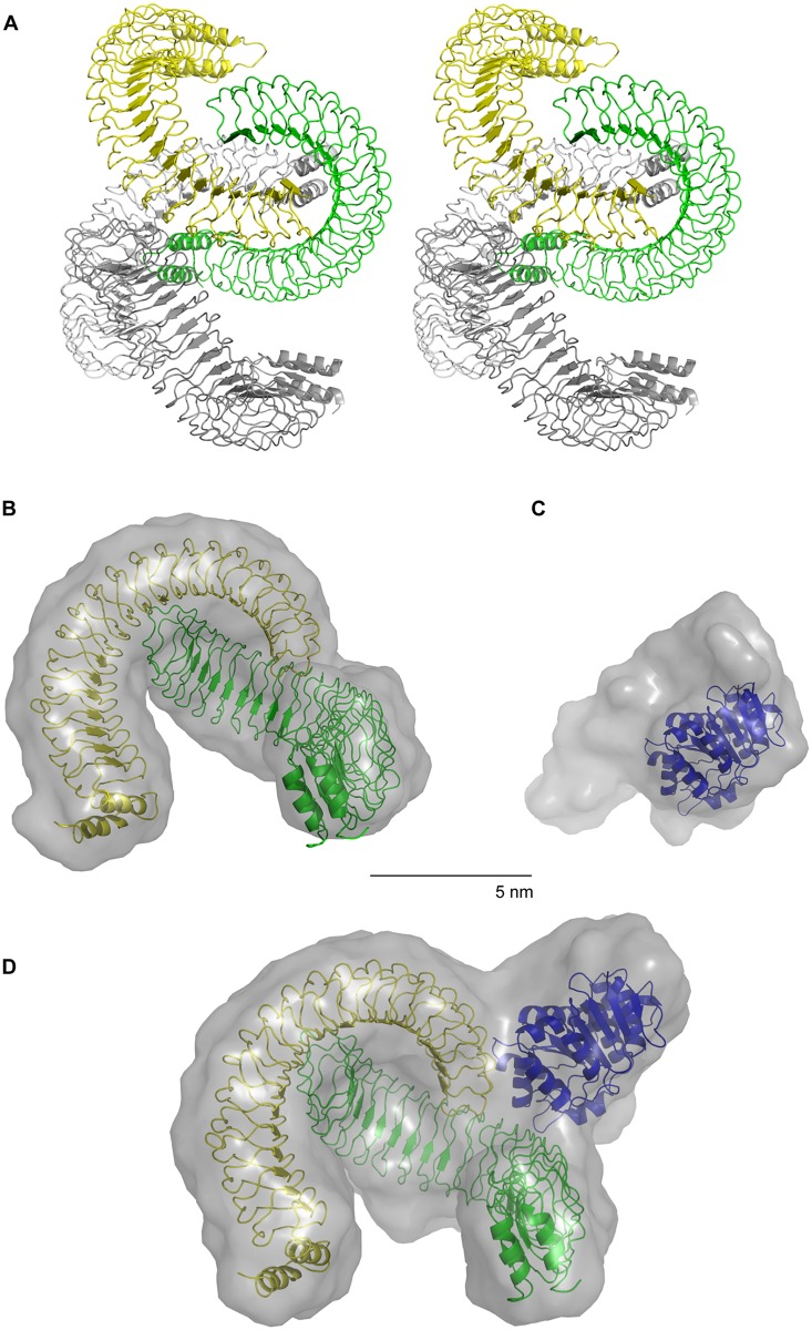
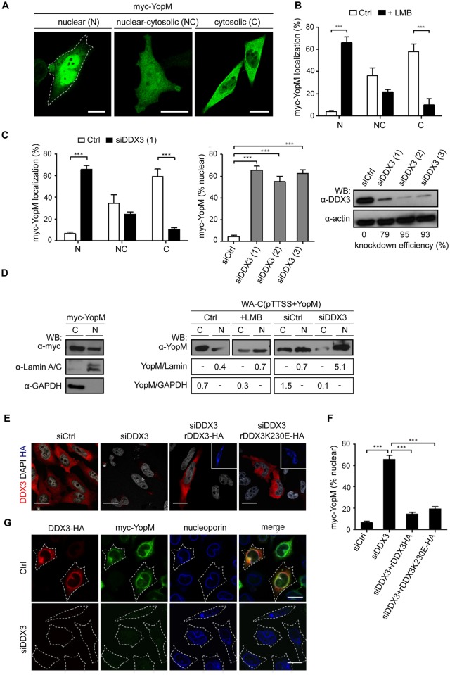
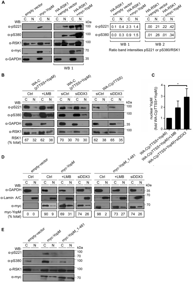
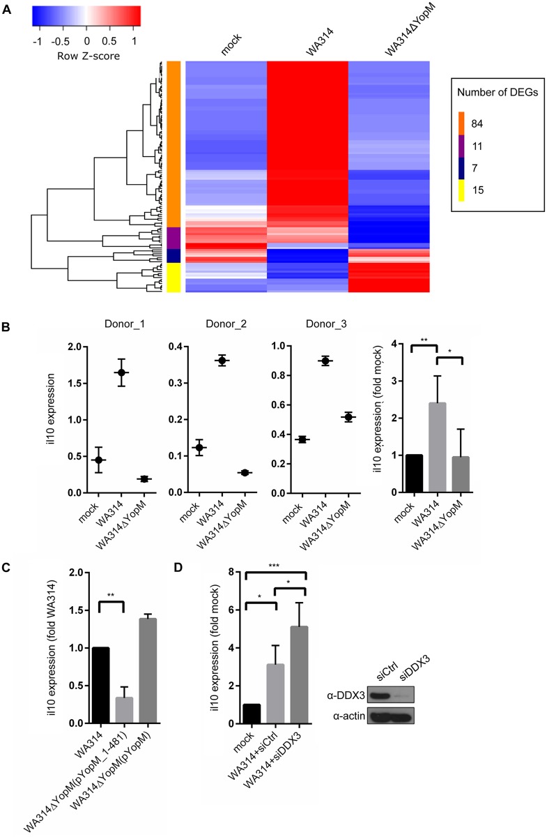
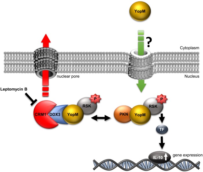
Yersinia outer protein M (YopM) is a crucial immunosuppressive effector of the plaque agent Yersinia pestis and other pathogenic Yersinia species. YopM enters the nucleus of host cells but neither the mechanisms governing its nucleocytoplasmic shuttling nor its intranuclear activities are known. Here we identify the DEAD-box helicase 3 (DDX3) as a novel interaction partner of Y. enterocolitica YopM and present the three-dimensional structure of a YopM:DDX3 complex. Knockdown of DDX3 or inhibition of the exportin chromosomal maintenance 1 (CRM1) increased the nuclear level of YopM suggesting that YopM exploits DDX3 to exit the nucleus via the CRM1 export pathway. Increased nuclear YopM levels caused enhanced phosphorylation of Ribosomal S6 Kinase 1 (RSK1) in the nucleus. In Y. enterocolitica infected primary human macrophages YopM increased the level of Interleukin-10 (IL-10) mRNA and this effect required interaction of YopM with RSK and was enhanced by blocking YopM's nuclear export. We propose that the DDX3/CRM1 mediated nucleocytoplasmic shuttling of YopM determines the extent of phosphorylation of RSK in the nucleus to control transcription of immunosuppressive cytokines.


Conflict of interest statement

The authors have declared that no competing interests exist.

Figures

Fig 1
Fig 1. Y. enterocolitica YopM interacts with DDX3 in host cells.
A) Coprecipitation of bacterially translocated YopM-SBP-CBP with DDX3, RSK1 and PKN in J774A.1 cells. J774A.1 cells were infected with WA314ΔYopM(pYopM-SBP-CBP), WA314ΔYopM(pYopM) or WA314ΔYopE(pYopE-SBP-CBP). Proteins eluting consecutively from streptavidin-sepharose (biotin elution) and calmodulin-sepharose (boiling in sample buffer) were analyzed by Western blot using indicated antibodies. B) GST-YopM pull-down of endogenous DDX3. GST-myc-YopM or GST expressed in HEK293T cells were precipitated with glutathione sepharose beads and analyzed by Western blot using indicated antibodies. C) Myc-YopM co-immunoprecipitates with endogenous DDX3. Endogenous DDX3 was immunoprecipitated in HEK293T cells expressing myc-YopM. Precipitates and whole cell lysates (WCL) were analyzed by Western blot using indicated antibodies. D) Bacterially translocated YopM co-immunoprecipitates with endogenous DDX3. Endogenous DDX3 was immunoprecipitated in HEK293T cells infected with WA314ΔYopM(pYopM) or WA314ΔYopM for 90 min. Precipitates and whole cell lysates (WCL) were analyzed by Western blot using indicated antibodies. E) DDX3 and PKN mutually exclude each other in RSK1/YopM containing complexes. HEK293T cells expressing either DDX3-FLAG (left panel) or PKN-FLAG (right panel) and where indicated myc-YopM were lysed and subjected to anti-FLAG immunoprecipitation. Precipitates and whole cell lysates (WCL) were analyzed by Western blot using indicated antibodies. In DDX3-FLAG immunoprecipitates, myc-YopM, RSK1 but no PKN and in PKN-FLAG immunoprecipitates myc-YopM, RSK1 but no DDX3 were detected.
Fig 2
Fig 2. YopM and DDX3 form a 2:1 molecular complex in solution.
A) Schematic representation of YopM from Y. enterocolitica WA314. The N-terminal blue rectangles in the YopM scheme correspond to the two α-helices (α1 and α2). LRRs are represented by numbered boxes. YopM_34–481 represents the crystallized variant of YopM (see below) containing all structured features of the protein. B) Schematic representation of DDX3. The gray rectangles in the DDX3 scheme correspond to the two helicase core domains (1 and 2). Truncated constructs used in GST-pulldown and immunoprecipitation are marked by amino acid numbers and brackets. C) GST-YopM pulldown of transfected DDX3 constructs. GST-Myc-YopM, GST and indicated HA-tagged DDX3 constructs were coexpressed in HEK293T cells and proteins precipitated by glutathione sepharose beads or in input (1/10 volume) were detected by Western blot using indicated antibodies. D) GST-YopM pulldown of bacterially expressed DDX3 constructs. Recombinant GST-YopM or GST bound to glutathione sepharose beads were incubated with bacterial lysates containing indicated His-tagged DDX3 constructs. DDX3 proteins precipitated by GST-YopM or in 1/10 lysate input were detected by Western blot using HisProbe-HRP conjugate. Input of GST or GST-YopM is shown in Coomassie stained gels. E) Microscale thermophoresis determines the binding affinity between YopM and DDX3. Binding strength of DDX3_1–418 to either YopM or YopM_34–481 was quantified by microscale thermophoresis as in Methods. The dissociation constants (Kd) for the respective interactions are indicated (mean +/- SD; one representative experiment out of three similar ones). For microscale thermophoresis experiment determining the binding strength of YopM to either DDX3_1–418 or DDX3_51–418 see also S1A Fig. F) Analytical size exclusion chromatography of the YopM:DDX3 complex. YopM, DDX3_1–418 or a 1:1 (molar ratio) mixture of both proteins were each run on a Superdex 200 gel filtration column. Molecular weights of the peak materials of the three runs (color coded and super-imposed in the figure) were calculated by comparing peak elution fractions with those of standard molecular weight marker proteins (S1B Fig for calibration curves, left panel). Indicated color coded peak fractions were analyzed by SDS PAGE. See also S1C Fig for analytical size exclusion chromatography of the YopM_34–481 and DDX3 1–418 complex. Corresponding calibration curve is shown in S1B Fig (right panel).
Fig 3
Fig 3. Crystal- and solution structure of YopM_34–481 and solution structures of DDX3_51–418 and the YopM_34-481/DDX3_51–418 complex.
A) YopM_34–481 was crystallized and its structure was solved (Methods; S2 Table). Stereo view shows ribbon representation of one asymmetric unit of the YopM_34–481 crystal (PDB code 4OW2). The asymmetric unit contains four molecules equivalent to two biological assemblies each represented by a dimer. YopM molecules of one dimer are colored in yellow and green and of the other dimer in light grey and grey. (B-D) The YopM_34–481 dimer, the DDX3_51–418 construct and the YopM_34-481/DDX3_51–418 complex were analyzed by small angle X-ray scattering (SAXS). B) The SASREF-model (Methods) of YopM_34–481 shown as transparent grey surface representation and the crystal structure of the YopM_34–481 dimer in ribbon representation are superimposed. C) The SASREF-model of DDX3_51–418 shown as transparent grey surface representation and the crystal structure of the ATPase domain of DDX3 (residues 167–418, PDB code 2I4I) are superimposed. D) The SASREF-model of the YopM_34-481/DDX3_51–418 complex shown as transparent grey surface representation is superimposed with the crystal structures of DDX3 (residues 167–418; [31] and the YopM_34–481 dimer (as presented in A). For experimental SAXS data see S2 Fig and S3 Table.
Fig 4
Fig 4. DDX3 mediates nuclear export of YopM via the CRM1 pathway.
A) Defining three patterns of nuclear/cytosolic distribution of YopM. HeLa cells expressing myc-YopM were immunofluorescence stained using anti-myc antibody and a nuclear (N), nuclear/cytosolic (NC) and cytosolic (C) distribution pattern of YopM was identified. Scale bar, 20μm. B) CRM1 inhibitor Leptomycin B increases percentage of nuclear localized YopM. HeLa cells expressing myc-YopM were not treated (Ctrl) or treated with 25 nM LMB for 4 h and then anti-myc immunofluorescence stained. The percentage of cells showing a N-, NC- or C localization of myc-YopM was determined. Each bar represents mean ± SED of 100 cells from three different experiments; ***p<0.001. For distribution of DDX3 within cells under LMB treatment see S3 Fig. C) Knockdown of DDX3 increases percentage of nuclear localized YopM. HeLa cells were treated with control siRNA (siCtrl) or DDX3 siRNA (siDDX3 No. 1, 2 or 3 as indicated) for 48 h, transfected with a vector encoding myc-YopM for 18 h and the percentage of cells displaying a N-, NC-, or C localization (left panel) or a N-localization (middle panel) of YopM was determined employing anti-myc immunofluorescence staining. Lysates of siRNA treated cells were analyzed by Western blot using anti-DDX3 antibody to determine knockdown efficiencies. Anti-actin antibody was used to verify equal loading of the SDS PAGE gels (right panel). Each bar represents mean ± SEM of 100 evaluated cells from 2–4 different experiments; ***p<0.001. D) Inhibition of CRM1 and knockdown of DDX3 increase the amount of YopM in nuclear cell fractions. (Left panel) Cytosolic (C) and nuclear (N) fractions of HEK293T cells expressing myc-YopM were prepared as in Methods and subjected to anti-myc immunoblot. Purity of fractions was verified using anti-GAPDH (cytosolic marker) and anti-Lamin A/C- antibody (nuclear marker), respectively. (Right panel) HEK293T cells were not treated (Ctrl) or treated with LMB, non targeting siRNA (siCtrl) or DDX3 siRNA (No. 3) and then infected with WA-C(pTTSS+YopM) for 1.5 h. Cytosolic (C) and nuclear (N) fractions of the cells were subjected to anti-YopM, anti-Lamin A/C and anti-GAPDH immunoblot. Band intensity ratios of nuclear YopM/Lamin and cytosolic YopM/GAPDH are depicted. E) Recomplementation of DDX3 knockdown cells with different DDX3 constructs. HeLa cells were treated with control siRNA (siCtrl) or DDX3 siRNA (siDDX3 No. 3) for 48 h and then transfected with vectors encoding siRNA-resistent HA-labelled DDX3 (rDDX3-HA) or the K230E mutant thereof (rDDX3K230E-HA). Cells were double immunofluorescence stained with anti-DDX3- and anti-HA antibodies and costained with DAPI for visualizing nuclei. Cells (re)expressing rDDX3 or rDDX3K230E stained positive with both, anti-DDX3- (red) and anti-HA (blue; insets) antibodies. Scale bar, 20 μm. F) Recomplementation of DDX3 reverses nuclear accumulation of YopM. HeLa cells were treated as in E) and were additionally transfected with myc-YopM and then immunofluorescence stained with anti-DDX3-, anti-HA and anti-myc antibodies. N-localized YopM was evaluated in cells either negative for DDX3 staining (siDDX3) or positive for DDX3 staining (remaining conditions). Each bar represents mean ± SEM of 100 cells from three different experiments; ***p<0.001. G) YopM and DDX3 colocalize at the outer nuclear membrane. HeLa cells were not treated (upper row) or treated with DDX3 siRNA No. 1 for 48 h (lower row) and then transfected with vectors encoding myc-YopM and DDX3-HA. Cells were permeablized with digitonin for 2 min before fixation to release the cytoplasm but leave the outer nuclear membrane intact and then stained with anti-HA- (red), anti-myc- (green) and anti-nucleoporin (blue) antibodies. Representative images are shown. Scale bar, 20 μm.
Fig 5
Fig 5. Nuclear YopM controls phosphorylation of nuclear RSK1.
A) Coexpression of HA-RSK1 and myc-YopM increases phosphorylation of nuclear RSK1. (Left panel) HEK293T cells were transfected with indicated expression vectors and cytosolic- (C) and nuclear (N) fractions were investigated by immunoblot using indicated antibodies. Anti-pS221- and Anti-pS380 antibodies detect the respective phosphorylated amino acids in RSK1. Exposure time of the anti-RSK1 panel in the HA-RSK1 expressing cells (right, WB 1) is about 1/5 of that of the respective panel in control cells (left). (Right panel) Intensities of the pS221- pS380- and RSK1 protein bands from WB 1 and WB 2, the latter derived from a second identical experiment were quantified and intensity ratios of pS221/RSK1 and pS380/RSK1 were calculated. B) Leptomycin B treatment and DDX3 knockdown increase phosphorylation of nuclear RSK1. (Left panel) HEK293T cells were not treated (Ctrl) or treated with 25 nM LMB for 4 h. (middle and right panels) HEK293T cells were treated with control siRNA (siCtrl) or DDX3 siRNA (siRNA No. 3) for 24 h. Cells were infected with Yersinia WA-C(pTTSS+YopM) (middle panel) or WA-C(pTTSS) (right panel) for 90 min and cytosolic- (C) and nuclear (N) fractions were investigated by immunoblot using indicated antibodies. Bottom panel shows percentage of total RSK1 present in the C- and N fraction of cells in each condition. C) Leptomycin B treatment and DDX3 knockdown increases nuclear YopM. HEK293T cells were not treated or treated with 25 nM LMB for 4 h or DDX3 siRNA (No. 3) for 24 h, infected with Yersinia WA-C(pTTSS+YopM) for 90 min and nuclear fractions of cells were investigated by anti-YopM immunoblot. Band intensities of YopM signals were determined by ImageJ. Each bar represents mean ± SEM of values from 4 experiments for LMB treatment and 3 experiments for DDX3 siRNA treatment; *p<0.05 (Bonferroni's multiple comparisons test). D) YopM_1–481 mutant defective in interaction with RSK accumulates in the nucleus and is exported from the nucleus in a LMB- and DDX3 dependent fashion. HEK293T cells were not treated (Ctrl), treated with 25 nM LMB for 4 h or DDX3 siRNA (siRNA No. 3) for 24 h and transfected with indicated expression vectors. Cytosolic- (C) and nuclear (N) fractions of cells in each condition were investigated by immunoblot using indicated antibodies. Bottom panel shows percentage of total myc-YopM present in the C- and N fraction of cells in the respective condition. See also S4 Fig for immunoprecipitaton of myc-YopM; myc-YopM_1–481 and myc-YopM_34–481 to verify the incapability of YopM lacking the C-terminus to bind RSK. E) YopM_1–481 mutant is unable to stimulate phosphorylation of RSK. Indicated myc-YopM constructs or empty vector were expressed in HEK293T cells and cytosolic (C) or nuclear (N) fractions were analyzed by Western blot using indicated antibodies.
Fig 6
Fig 6. YopM upregulates IL-10 expression in Yersinia infected human macrophages.
A) Heatmap of expression values of differentially expressed genes (DEGs) in human macrophages not infected (mock) or infected with WA314 or WA314ΔYopM. Duplicates (two different donors) of primary human macrophages were infected without (mock) or with Yersinia enterocolitica WA314 or WA314ΔYopM for 6 h. Total RNA was prepared from each sample and subjected to RNA-seq. The scaled expression of each set of replicates, denoted as the row Z-score, is plotted in a red-blue color scale. Red indicates high and blue indicates low expression. Only genes with an absolute log2-fold change greater or equal 2 and p-value smaller or equal 0.01 are shown. These genes were hierarchically clustered (complete linkage) according to their expression profiles. The resulting distinct sets of genes are indicated by the orange-, purple-, blue- and yellow color bar. The number of DEGs in each cluster is indicated. B) Analysis of IL-10 expression in Yersinia infected human macrophages from different donors. Total RNA was isolated from primary human macrophages that were mock infected or infected with WA314 or WA314ΔYopM for 6 h. The RNA was subjected to quantitative RT-PCR using human IL-10 specific primers. IL-10 expression was normalized to expression of three housekeeping genes (GAPDH, TBP, B2M). For each condition triplicate samples of macrophages derived from seven different donors (Donor_1 to Donor_7) were investigated (data from Donor_4 to Donor_7 in S5 Fig). Each bar in graph represents mean ± SD of values from all 7 donors; **p<0.01, *p<0.05. C) Induction of IL-10 expression requires interaction of YopM with RSK. Experimental procedures as in B) with the difference that macrophages were infected with WA314ΔYopM(pYopM_1–481) and WA314ΔYopM(pYopM). Each Bar in graph represents mean ± SD of values from 3 different donors; **p<0.01. D) DDX3 knockdown increases IL-10 expression in Yersinia infected human macrophages. Primary human macrophages were transfected with control siRNA (mock, siCtrl) or DDX3 siRNA (siDDX3 No. 3) for 72 h and not infected (mock) or infected with WA314 for 6 h. Total RNA was subjected to quantitative RT-PCR as in B). Each Bar in graph represents mean ± SD of values from 4 different donors; *p<0.05, ***p<0.001. Western blot verifies DDX3 knockdown in the macrophages.
Fig 7
Fig 7. Model of nuclear shuttling and intranuclear activity of YopM.
YopM enters the nucleus from the cytoplasm of infected cells through an unknown pathway. In the nucleus YopM forms at least two complexes: YopM:RSK:PKN and YopM:RSK:DDX3. Hyperphosphorylated RSK within the complex induces IL-10 expression. DDX3 mediates nuclear export of YopM via the LMB-sensitive CRM1 export pathway. Abbreviations: RSK, Ribosomal S6 kinase; PKN, Protein kinase N; DDX3, DEAD box helicase 3; CRM1, chromosome region maintenance 1; P, phosphate group; TF, transcription factor.

References

    1. Leung KY, Straley SC. The yopM gene of Yersinia pestis encodes a released protein having homology with the human platelet surface protein GPIb alpha. Journal of bacteriology. 1989;171(9):4623–32. - PMC - PubMed
    1. Boland A, Havaux S, Cornelis GR. Heterogeneity of the Yersinia YopM protein. Microbial pathogenesis. 1998;25(6):343–8. - PubMed
    1. Boland A, Sory MP, Iriarte M, Kerbourch C, Wattiau P, Cornelis GR. Status of YopM and YopN in the Yersinia Yop virulon: YopM of Y.enterocolitica is internalized inside the cytosol of PU5-1.8 macrophages by the YopB, D, N delivery apparatus. The EMBO journal. 1996;15(19):5191–201. - PMC - PubMed
    1. Uittenbogaard AM, Chelvarajan RL, Myers-Morales T, Gorman AA, Brickey WJ, Ye Z, et al. Toward a molecular pathogenic pathway for Yersinia pestis YopM. Frontiers in cellular and infection microbiology. 2012;2:155 10.3389/fcimb.2012.00155 - DOI - PMC - PubMed
    1. Galan JE, Wolf-Watz H. Protein delivery into eukaryotic cells by type III secretion machines. Nature. 2006;444(7119):567–73. - PubMed

MeSH terms