

Modulation of the Mesenchymal Stem Cell Secretome Using Computer-Controlled Bioreactors: Impact on Neuronal Cell Proliferation, Survival and Differentiation
- PMID: 27301770
- PMCID: PMC4908397
- DOI: 10.1038/srep27791
Modulation of the Mesenchymal Stem Cell Secretome Using Computer-Controlled Bioreactors: Impact on Neuronal Cell Proliferation, Survival and Differentiation
Abstract
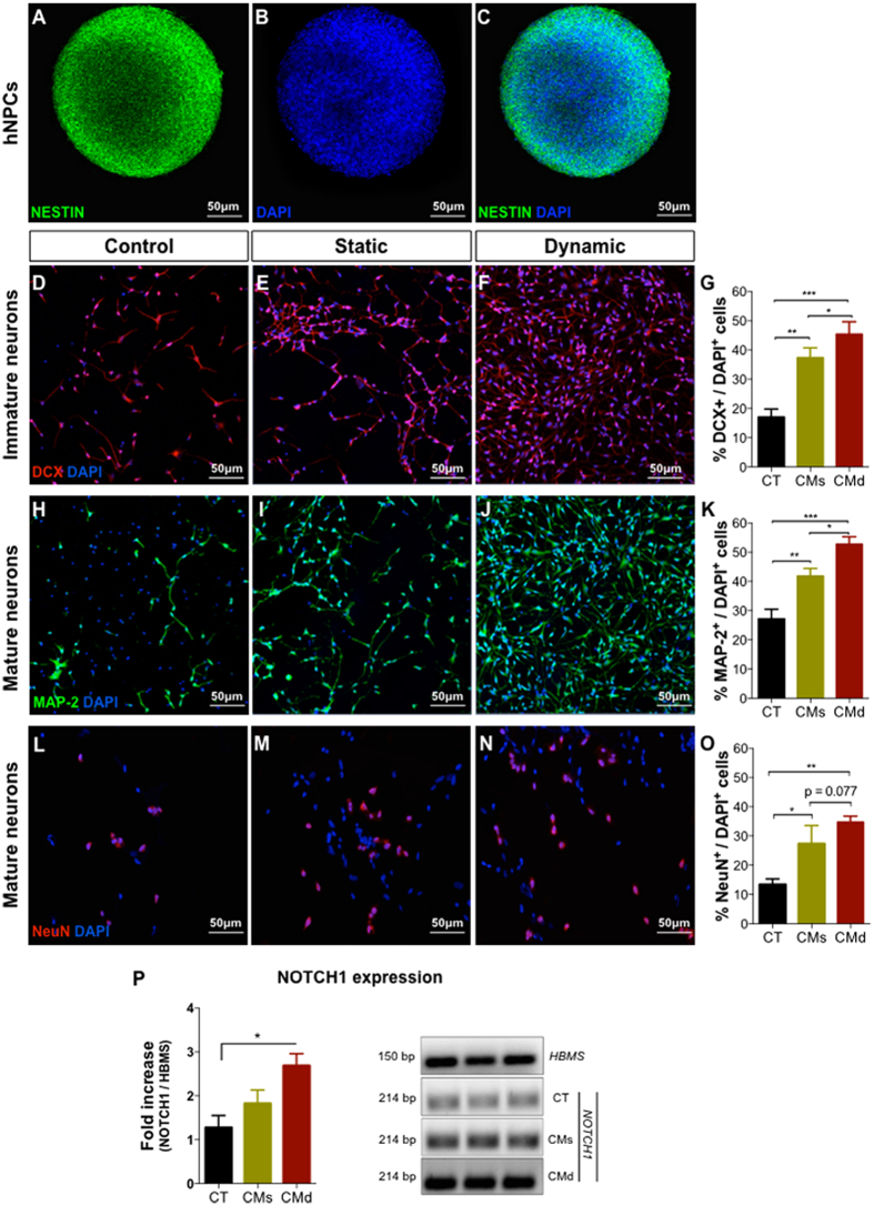
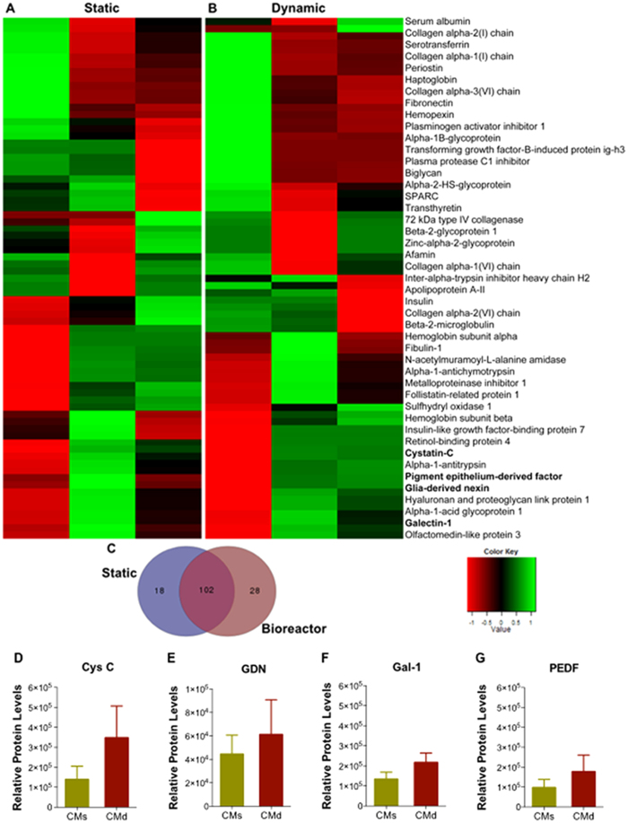
In recent years it has been shown that the therapeutic benefits of human mesenchymal stem/stromal cells (hMSCs) in the Central Nervous System (CNS) are mainly attributed to their secretome. The implementation of computer-controlled suspension bioreactors has shown to be a viable route for the expansion of these cells to large numbers. As hMSCs actively respond to their culture environment, there is the hypothesis that one can modulate its secretome through their use. Herein, we present data indicating that the use of computer-controlled suspension bioreactors enhanced the neuroregulatory profile of hMSCs secretome. Indeed, higher levels of in vitro neuronal differentiation and NOTCH1 expression in human neural progenitor cells (hNPCs) were observed when these cells were incubated with the secretome of dynamically cultured hMSCs. A similar trend was also observed in the hippocampal dentate gyrus (DG) of rat brains where, upon injection, an enhanced neuronal and astrocytic survival and differentiation, was observed. Proteomic analysis also revealed that the dynamic culturing of hMSCs increased the secretion of several neuroregulatory molecules and miRNAs present in hMSCs secretome. In summary, the appropriate use of dynamic culture conditions can represent an important asset for the development of future neuro-regenerative strategies involving the use of hMSCs secretome.
Figures


Similar articles
-
Unveiling the Differences of Secretome of Human Bone Marrow Mesenchymal Stem Cells, Adipose Tissue-Derived Stem Cells, and Human Umbilical Cord Perivascular Cells: A Proteomic Analysis.Stem Cells Dev. 2016 Jul 15;25(14):1073-83. doi: 10.1089/scd.2016.0048. Epub 2016 Jun 27. Stem Cells Dev. 2016. PMID: 27226274
-
Tips on How to Collect and Administer the Mesenchymal Stem Cell Secretome for Central Nervous System Applications.Methods Mol Biol. 2016;1416:457-65. doi: 10.1007/978-1-4939-3584-0_27. Methods Mol Biol. 2016. PMID: 27236689
-
Do hypoxia/normoxia culturing conditions change the neuroregulatory profile of Wharton Jelly mesenchymal stem cell secretome?Stem Cell Res Ther. 2015 Jul 24;6(1):133. doi: 10.1186/s13287-015-0124-z. Stem Cell Res Ther. 2015. PMID: 26204925 Free PMC article.
-
Proteomic techniques for characterisation of mesenchymal stem cell secretome.Biochimie. 2013 Dec;95(12):2196-211. doi: 10.1016/j.biochi.2013.07.015. Epub 2013 Jul 20. Biochimie. 2013. PMID: 23880644 Review.
-
Stem cells: insights into the secretome.Biochim Biophys Acta. 2013 Nov;1834(11):2380-4. doi: 10.1016/j.bbapap.2013.01.032. Epub 2013 Jan 31. Biochim Biophys Acta. 2013. PMID: 23376432 Review.
Cited by
-
Secretome from In Vitro Mechanically Loaded Myoblasts Induces Tenocyte Migration, Transition to a Fibroblastic Phenotype and Suppression of Collagen Production.Int J Mol Sci. 2021 Dec 3;22(23):13089. doi: 10.3390/ijms222313089. Int J Mol Sci. 2021. PMID: 34884895 Free PMC article.
-
Biological Considerations in Scaling Up Therapeutic Cell Manufacturing.Front Pharmacol. 2020 May 13;11:654. doi: 10.3389/fphar.2020.00654. eCollection 2020. Front Pharmacol. 2020. PMID: 32528277 Free PMC article. Review.
-
Three-Dimensional Bioreactor Technologies for the Cocultivation of Human Mesenchymal Stem/Stromal Cells and Beta Cells.Stem Cells Int. 2018 Mar 14;2018:2547098. doi: 10.1155/2018/2547098. eCollection 2018. Stem Cells Int. 2018. PMID: 29731775 Free PMC article. Review.
-
Mesenchymal stromal cell-derived secretome-based therapy for neurodegenerative diseases: overview of clinical trials.Stem Cell Res Ther. 2023 May 4;14(1):122. doi: 10.1186/s13287-023-03264-0. Stem Cell Res Ther. 2023. PMID: 37143147 Free PMC article. Review.
-
Bioprocessing of Mesenchymal Stem Cells and Their Derivatives: Toward Cell-Free Therapeutics.Stem Cells Int. 2018 Sep 12;2018:9415367. doi: 10.1155/2018/9415367. eCollection 2018. Stem Cells Int. 2018. PMID: 30275839 Free PMC article. Review.
References
-
- Salgado A. J. & Gimble J. M. Secretome of mesenchymal stem/stromal cells in regenerative medicine. Biochimie 95, 2195 (2013). - PubMed
-
- Arslan F. et al.. Mesenchymal stem cell-derived exosomes increase ATP levels, decrease oxidative stress and activate PI3K/Akt pathway to enhance myocardial viability and prevent adverse remodeling after myocardial ischemia/reperfusion injury. Stem Cell Res 10, 301–312 (2013). - PubMed
Publication types
MeSH terms
Substances
LinkOut - more resources
Full Text Sources
Other Literature Sources
Medical
Research Materials
