. 2017 Jan;19(1):e12628.
doi: 10.1111/cmi.12628. Epub 2016 Jul 8.

Arabidopsis late blight: infection of a nonhost plant by Albugo laibachii enables full colonization by Phytophthora infestans

Affiliations

Arabidopsis late blight: infection of a nonhost plant by Albugo laibachii enables full colonization by Phytophthora infestans

Khaoula Belhaj et al. Cell Microbiol. 2017 Jan.

Abstract

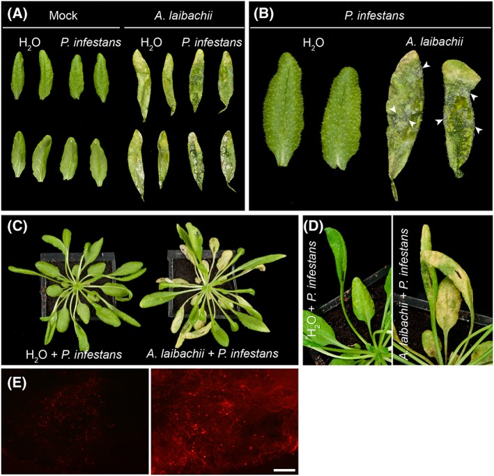
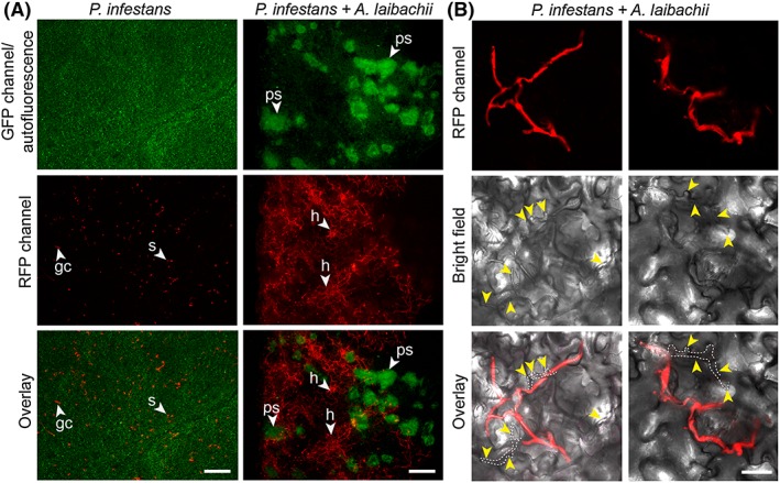
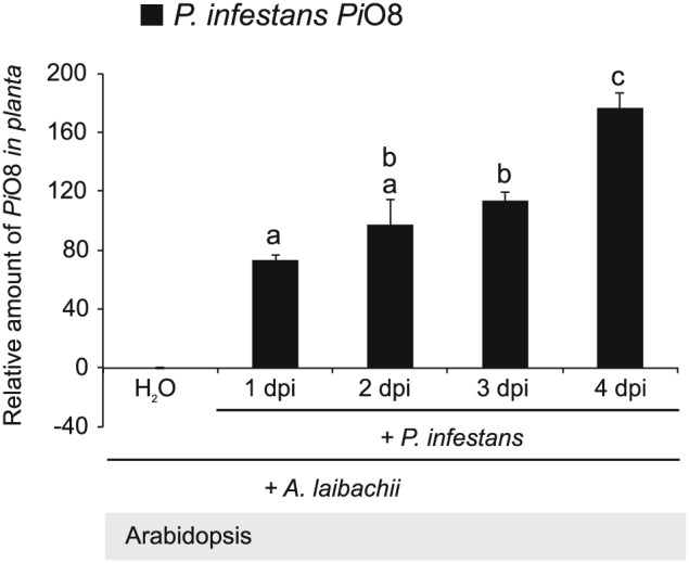
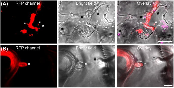
The oomycete pathogen Phytophthora infestans causes potato late blight, and as a potato and tomato specialist pathogen, is seemingly poorly adapted to infect plants outside the Solanaceae. Here, we report the unexpected finding that P. infestans can infect Arabidopsis thaliana when another oomycete pathogen, Albugo laibachii, has colonized the host plant. The behaviour and speed of P. infestans infection in Arabidopsis pre-infected with A. laibachii resemble P. infestans infection of susceptible potato plants. Transcriptional profiling of P. infestans genes during infection revealed a significant overlap in the sets of secreted-protein genes that are induced in P. infestans upon colonization of potato and susceptible Arabidopsis, suggesting major similarities in P. infestans gene expression dynamics on the two plant species. Furthermore, we found haustoria of A. laibachii and P. infestans within the same Arabidopsis cells. This Arabidopsis-A. laibachii-P. infestans tripartite interaction opens up various possibilities to dissect the molecular mechanisms of P. infestans infection and the processes occurring in co-infected Arabidopsis cells.


Figures

Figure 1
Figure 1
Albugo laibachii enables Phytophthora infestans to colonize Arabidopsis on detached leaves and on whole plants. A. Control leaves (Mock) or leaves from A. thaliana Col‐0 plants pre‐infected with A. laibachii were detached and droplets of water (H20) or P. infestans spore solution were applied to their abaxial sides and incubated for 4 days in high humidity. B. A close‐up of (A) reveals P. infestans sporulation (arrowheads) as a dense cover of leaves pre‐infected by A. laibachii only. B. Albugo laibachii enables P. infestans to colonize leaves infected on a whole plant. A. thaliana Col‐0 plants treated with water (H20) or pre‐infected with A. laibachii were inoculated with droplets of water (H20) or P. infestans spore solution and incubated in high humidity. Macroscopic observations of disease symptoms on whole plants at 3 days post inoculation. D. A close‐up of (C) reveals P. infestans disease symptoms only on leaves pre‐colonized by A. laibachii (right panel). E. The extent of P. infestans 88069td hyphal colonization (under RFP illumination) was assessed 3 days post inoculation using epifluorescence microscopy. Scale bar = 250 µm. All experiments were performed at least twice with similar results.
Figure 2
Figure 2
A. laibachii pre‐infection supports extensive hyphal growth of P. infestans in Arabidopsis. A. Abaxial sides of control leaves of A. thaliana Col‐0 (left column) and leaves pre‐infected with A. laibachii (right column) have been infected with red fluorescent P. infestans 88069td. The extent of A. laibachii sporulation (under GFP illumination visible as green autofluorescent pustules, upper row) and P. infestans hyphal colonization (visible under RFP illumination as red hyphal network, middle row) was assessed 3 days post inoculation using epifluorescence microscopy. Bottom row represents merged fluorescence pictures. B. Abaxial sides of co‐infected leaves at 2 dpi exhibiting dual colonization of P. infestans (in red) and by A. laibachii hyphae (not fluorescently labelled, indicated by yellow arrowheads) within the same area. All experiments were performed twice with similar results. Abbreviations: ps: pustules, h: hyphae; s: spores, gc: germinating cyst. Scale bars = 250 µm (A) or 50 µm (B).
Figure 3
Figure 3
Quantification of P. infestans biomass upon infection of A. thaliana pre‐infected with A. laibachii. Five‐week old leaves of A. thaliana Col‐0 pre‐infected with A. laibachii were detached and drop‐inoculated with a zoospore suspension of P. infestans isolate 06_3928A or mock‐treated with water applied to their abaxial sides and incubated for 4 days under high humidity. DNA was extracted at 0, 1, 2, 3, and 4 days post inoculation and used for quantitative PCR (qPCR) for PiO8 with gene‐specific primers for P. infestans. Pathogen DNA levels were normalized to the Arabidopsis SAND gene (At2g28390) and the relative amount of Pi08 was normalized to the DNA level in mock‐inoculated samples. Data are representative of one biological replicate with three technical replicates of qPCR reaction. Bars represent ratio between mean normalized expression of the infected samples with P. infestans and A. laibachii and the mock‐treated sample with A. laibachii and water (calibrator) (Mean ± SE). Data was analysed using repeated measures two way‐ANOVA (P < 0.0001). Letters indicate significant results of Fisher's LSD post‐hoc test. Experiment was performed twice with similar results.
Figure 4
Figure 4
Hypersusceptibility of A. thaliana to P. infestans in leaves pre‐infected with A. laibachii is accompanied by a loss of the hypersensitive response Five‐week‐old leaves of A. thaliana Col‐0 mock‐treated or pre‐infected with A. laibachii were drop‐inoculated with a zoospore suspension of red fluorescent P. infestans 88069td Pathogen structures and autofluorescent dead epidermal cells were visualized with confocal laser scanning microscopy at 16 hpi (A–D) and at 24 hpi (E–H) in samples treated with P. infestans only (A, C, E, F) and in co‐infection experiments with A. laibachii (B, D, G, H). Panel F represents a maximum projection of images produced from 18 Z stacks showing a hypersensitive response of the same area as panel E. All experiments were performed twice with similar results. (I) Counts of dead cells per leaf after infection with P. infestans in the presence or absence of pre‐infection with A. laibachii. Data are representative of two biological replicates. Each replicate consists of counts from eight independent leaves. Bars represent mean ± SD. Data was analysed using one‐way‐ANOVA (P < 0.0001). Letters indicate significant results of Fisher's LSD post‐hoc test. The two light microscopy inserts show examples of an HR cell death in infected leaves with P. infestans only (top panel) and of absence of HR cell death in co‐infection experiments with A. laibachii and P. infestans (low panel) at 24 hpi. Abbreviations: sp: spores, gt: germ tube, ap: appressorium, ec: empty cyst, pp: penetration peg, iv: infection vesicle, HR: hypersensitive cell death, max. proj.: maximum projection. Scale bar = 25 µm (A–F) or 7.5 µm (G–H).
Figure 5
Figure 5
Phytophthora infestans and Albugo laibachii can form haustoria in the same Arabidopsis cell. A. A. thaliana Col‐0 precolonized with A. laibachii was infected with red fluorescent P. infestans 88069td. Inspection by microscopy at 2 dpi revealed the presence of haustoria. B. Frequently, plant cells were observed to harbour digit‐like, red fluorescent P. infestans 88069td haustoria as well as knob‐like A. laibachii haustoria. All experiments were performed twice with similar results. Abbreviations: #: haustoria of A. laibachii, *: haustoria of P. infestans. Scale bar = 25 µm (A) or 7.5 µm (B).
Figure 6
Figure 6
Similar sets of effectors are induced during P. infestans colonization of potato (Solanum tuberosum) and Arabidopsis pre‐infected with A. laibachii. A. Numbers of commonly and uniquely induced genes encoding secreted P. infestans proteins and effectors (a subset of the secreted proteins). B. Dot blot comparing the transcript levels of P. infestans effector‐encoding genes between S. tuberosum and Arabidopsis pre‐infected with A. laibachii at 2 (left panel) and 3 days post infection (right panel). C. Gene expression intensities relative to the average expression intensity in media (Rye sucrose) are shown for genes encoding avirulence proteins (Gene IDs in parentheses) during the interaction of P. infestans with A. laibachii pre‐infected Arabidopsis leaves (left panel) and S. tuberosum leaves (right panel) induced at 2 dpi and 3 dpi. Genes encoding ubiquitin ligases, Elongation factor 2, and Actin are shown as uninduced controls. All expression intensities are log2 transformed.

References

    1. Agrios, G.N. (2005) Plant Pathology. San Diego, CA: Elsevier.
    1. Akrofi, A.Y. , Amoako‐Atta, I. , Assuah, M. , and Asare, E.K. (2015) Black pod disease on cacao (Theobroma cacao, L) in Ghana: spread of Phytophthora megakarya and role of economic plants in the disease epidemiology. Crop Prot 72: 66–75.
    1. Anders, S. , Pyl, P.T. , and Huber, W. (2015) HTSeq—a Python framework to work with high‐throughput sequencing data. Bioinformatics btu638 31: 166–169. - PMC - PubMed
    1. Ayliffe, M. , Jin, Y. , Kang, Z. , Persson, M. , Steffenson, B. , Wang, S. , and Leung, H. (2011) Determining the basis of nonhost resistance in rice to cereal rusts. Euphytica 179: 33–40.
    1. Bains, S. , and Jhooty, J. (1985) Association of Peronospora parasitica with Albugo candida on Brassica juncea leaves. J Phytopathol 112: 28–31.

Publication types

MeSH terms