Review
. 2016 Jun 15;143(12):2056-65.
doi: 10.1242/dev.127886.

Extracellular matrix motion and early morphogenesis

Affiliations
Review

Extracellular matrix motion and early morphogenesis

Rajprasad Loganathan et al. Development. .

Abstract

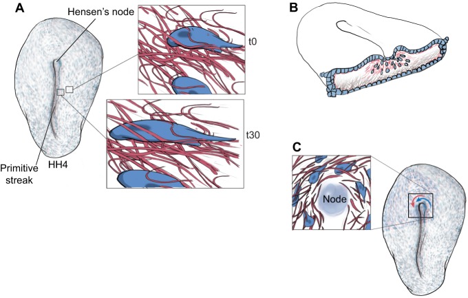
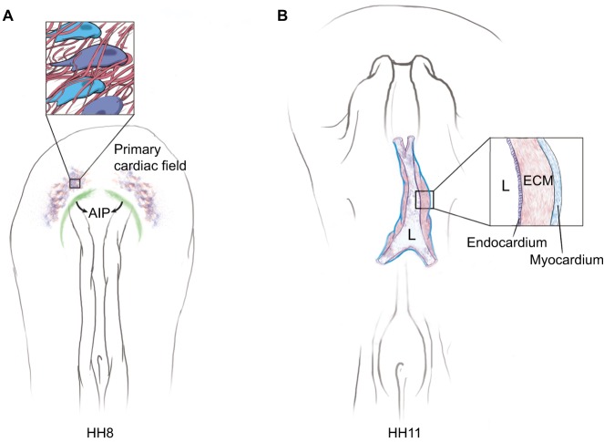
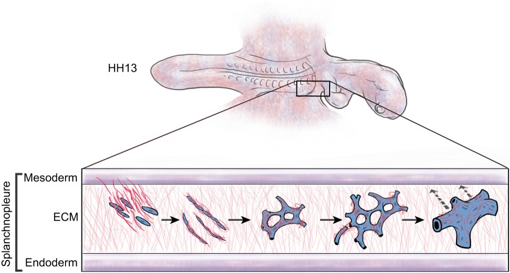
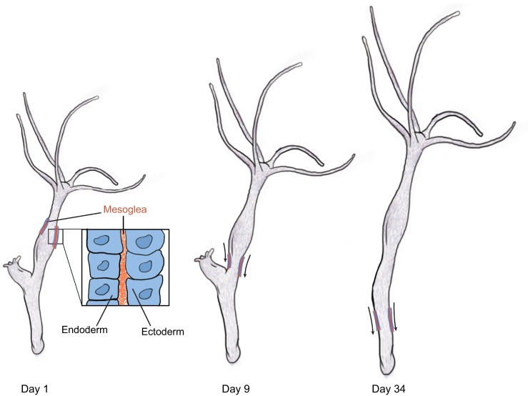
For over a century, embryologists who studied cellular motion in early amniotes generally assumed that morphogenetic movement reflected migration relative to a static extracellular matrix (ECM) scaffold. However, as we discuss in this Review, recent investigations reveal that the ECM is also moving during morphogenesis. Time-lapse studies show how convective tissue displacement patterns, as visualized by ECM markers, contribute to morphogenesis and organogenesis. Computational image analysis distinguishes between cell-autonomous (active) displacements and convection caused by large-scale (composite) tissue movements. Modern quantification of large-scale 'total' cellular motion and the accompanying ECM motion in the embryo demonstrates that a dynamic ECM is required for generation of the emergent motion patterns that drive amniote morphogenesis.

Keywords: Amniote morphogenesis; Emergent patterns; Extracellular matrix dynamics; Tissue-scale motion.


Conflict of interest statement

Competing interests

The authors declare no competing or financial interests.

Figures

Fig. 1.
Fig. 1.
Composite tissue (cells plus ECM) motion during early embryogenesis. (A) ECM motion is present in avian embryos as early as primitive streak formation. Time-lapse recordings of cells (blue) and ECM fibers (red) show both embryonic tissue components moving as a composite [compare insets at time (t) 0 and 30 min later], along with concomitant cell shape changes. (B) During gastrulation, ECM fibers (pink) move through the primitive streak (ingress) along with prospective mesendodermal cells (blue). See also Movie 1. (C) A third example of ECM fiber motion occurs near Hensen's node (the ‘organizer’ in birds). Nodal cells, adjacent epiblastic cells and ECM fibers move in unison (blue and red arrows). The organizer tissue undergoes rotational motion that determines left-right asymmetry with respect to the head-to-tail axis. Only counterclockwise motion is illustrated for simplicity; however, clockwise rotation of more remote epiblastic cells occurs in the peri-nodal region (see Cui et al., 2009b).
Fig. 2.
Fig. 2.
Tissue-scale motion dominates amniote heart formation. (A) Medial displacement of the primary cardiac field is driven by the centripetal forces (arrows) of anterior intestinal portal (AIP; green) regression. As the tissue moves the ECM fibers (red), endocardial progenitors (purple) and myocardial progenitors (blue) are propelled toward the midline (arrows). There is negligible independent cellular motility during AIP regression. See Movie 3 and Aleksandrova et al. (2012). (B) Driven by tissue deformation of cardiac progenitors and ECM, the right and left heart primordia fuse at the midline – the cardinal step in forming the tube-within-a-tube morphology of the amniote heart. Note that once fusion occurs, an expansive ECM (cardiac jelly) separates the myocardium and endocardium. Many of the ECM fibrils in the cardiac jelly (inset) were ‘born’ hours earlier and were subsequently transported into the tubular heart by the AIP-driven tissue deformation(s) (see Aleksandrova et al., 2015a). L, lumen of the primitive heart chamber.
Fig. 3.
Fig. 3.
Tissue-scale motion during primary vasculogenesis. ECM motion is an integral component of the multi-stage primary vasculogenic process. Collective motion (ECM plus endothelium) influences all stages of primary vascular network formation. The process takes place within a splanchnopleural ECM that is expanding in three dimensions. Primordial endothelial progenitor cells move ventrally from the splanchnic mesoderm into the ECM and begin extension and protrusive behavior. Local ‘active’ motion of primordial endothelial cells leads to multicellular vascular cords (no lumen) that are subject to large-scale tissue drift/expansion (cells and ECM). As the lateral embryonic plate expands, the vascular cords coalesce into a hexagonal pattern, driven by both cellular motility and tissue motion, resulting in a vascular network composed of relatively small-caliber tubules. Further pattern formation involves tissue-scale remodeling of polygonal vascular networks. Specifically, a complex tissue-driven process begins whereby the lumens of some small-caliber vessels are forced together and fuse their respective lumens. Simultaneously, pruning results in the loss of other endothelial tubes. The result is morphogenesis of the great vessels, such as the aortae and the omphalomesenteric veins [see Movie 4 and Sato et al. (2010)]. During vasculogenesis, wide-scale tissue drift draws the intraembryonic and extraembryonic primary vasculature toward the midline (dashed arrows). Convergence of the extraembryonic vasculature toward the intraembryonic vasculature enables docking with the aortae and omphalomesenteric veins, resulting in a functional circulatory network. In amniotes the majority of morphogenetic motion experienced by a given endothelial cell is propelled by tissue-level events; meanwhile, local motility, cell extension and protrusive activity contribute to local shaping of individual vessels. Primary vascular pattern formation is emergent; there is no evidence that the process is predetermined or genetically regulated (see Perryn et al., 2008).
Fig. 4.
Fig. 4.
Hydra epithelia and mesoglea move together. Schematic of an experiment from Aufschnaiter et al. (2011) in which a graft containing fluorescently labeled mesoglea (primitive ECM, orange) and epithelial cells (blue) is displaced along the body column toward the aboral end of a Hydra (arrows) over the course of several days after grafting. The mesoglea and the epithelium (inset) move as a cohesive unit, demonstrating very early evolutionary evidence of morphogenetic tissue motion; the process involves extant ECM molecules such as collagen and laminin. A daughter hydra was seen budding off from the mother during the course of this experiment (day 1 and day 9).

References

    1. Abercrombie M. (1977). Concepts in morphogenesis. Proc. R. Soc. B Biol. Sci. 199, 337-344. 10.1098/rspb.1977.0145 - DOI - PubMed
    1. Adams J. C. (2013). Extracellular matrix evolution: an overview. In Evolution of Extracellular Matrix (ed. Keeley F. W. and Mecham R. P.), pp. 1-25. Berlin: Springer.
    1. Aleksandrova A., Czirók A., Szabó A., Filla M. B., Hossain M. J., Whelan P. F., Lansford R. and Rongish B. J. (2012). Convective tissue movements play a major role in avian endocardial morphogenesis. Dev. Biol. 363, 348-361. 10.1016/j.ydbio.2011.12.036 - DOI - PMC - PubMed
    1. Aleksandrova A., Czirok A., Kosa E., Galkin O., Cheuvront T. J. and Rongish B. J. (2015a). The endoderm and myocardium join forces to drive early heart tube assembly. Dev. Biol. 404, 40-54. 10.1016/j.ydbio.2015.04.016 - DOI - PMC - PubMed
    1. Aleksandrova A., Rongish B. J., Little C. D. and Czirók A. (2015b). Active cell and ECM movements during development. Methods Mol. Biol. 1189, 123-132. 10.1007/978-1-4939-1164-6_9 - DOI - PMC - PubMed

Publication types

LinkOut - more resources