. 2016 Sep;283(Pt A):142-50.
doi: 10.1016/j.expneurol.2016.06.012. Epub 2016 Jun 11.

Deep brain stimulation improves behavior and modulates neural circuits in a rodent model of schizophrenia

Affiliations

Deep brain stimulation improves behavior and modulates neural circuits in a rodent model of schizophrenia

Lior Bikovsky et al. Exp Neurol. 2016 Sep.

Abstract

Schizophrenia is a debilitating psychiatric disorder with a significant number of patients not adequately responding to treatment. Deep brain stimulation (DBS) is a surgical technique currently investigated for medically-refractory psychiatric disorders. Here, we use the poly I:C rat model of schizophrenia to study the effects of medial prefrontal cortex (mPFC) and nucleus accumbens (Nacc) DBS on two behavioral schizophrenia-like deficits, i.e. sensorimotor gating, as reflected by disrupted prepulse inhibition (PPI), and attentional selectivity, as reflected by disrupted latent inhibition (LI). In addition, the neurocircuitry influenced by DBS was studied using FDG PET. We found that mPFC- and Nacc-DBS alleviated PPI and LI abnormalities in poly I:C offspring, whereas Nacc- but not mPFC-DBS disrupted PPI and LI in saline offspring. In saline offspring, mPFC-DBS increased metabolism in the parietal cortex, striatum, ventral hippocampus and Nacc, while reducing it in the brainstem, cerebellum, hypothalamus and periaqueductal gray. Nacc-DBS, on the other hand, increased activity in the ventral hippocampus and olfactory bulb and reduced it in the septal area, brainstem, periaqueductal gray and hypothalamus. In poly I:C offspring changes in metabolism following mPFC-DBS were similar to those recorded in saline offspring, except for a reduced activity in the brainstem and hypothalamus. In contrast, Nacc-DBS did not induce any statistical changes in brain metabolism in poly I:C offspring. Our study shows that mPFC- or Nacc-DBS delivered to the adult progeny of poly I:C treated dams improves deficits in PPI and LI. Despite common behavioral responses, stimulation in the two targets induced different metabolic effects.

Keywords: Deep brain stimulation; FDG-PET; Latent inhibition; Maternal immune activation rat model; Prepulse inhibition; Schizophrenia.


Figures

Fig. 1
Fig. 1
Electrode placements. (a) Schematic reconstruction of electrode placements in either the mPFC (left) or Nacc (right), histologically verified in rats undergoing PPI (grey circles) and LI (black circles: PE; white circles: NPE) circles) experiments. (b) Representative CT scan showing electrode positioned in either the mPFC (upper) or Nacc (bottom).
Fig. 2
Fig. 2
Effects of DBS on PPI with averaged PPI values across three different prepulse intensities in the offspring of saline and poly I:C treated rats: For each brain area (mPFC (a) and Nacc (b)), animals underwent three tests sessions 24 h apart. Session 1 (baseline 1) was conducted with no stimulation applied. Session 2 was conducted while animals were receiving active DBS. Session 3 (baseline 2) was carried out with DBS turned off to assess whether the observed effects in Session 2 were permanent or transient. DBS in either the mPFC or Nacc significantly decreased PPI deficits in poly I:C offspring. In controls, while no changes were observed after mPFC-DBS, stimulation of the Nacc has induced significant PPI deficits. (*) Significantly different from baseline 1 within groups. (§) Significant differences between groups (saline vs. poly I:C). Results are presented as means ± S.E.M.; * p < 0.05.
Fig. 3
Fig. 3
Effects of DBS on LI in the offspring of saline and poly I:C treated dams: (a) mPFC-DBS reversed prenatal poly I:C-induced deficits in LI without affecting LI in controls. (b) Nacc-DBS improved prenatal poly I:C-induced LI deficits while deteriorating this behavior in controls. Data represent mean ± S.E.M of log times to complete licks 76–100 (after tone onset) of pre-exposed (PE) and non-pre-exposed (NPE) offspring of saline- or poly I:C treated dams that did (DBS) or did not receive DBS (sham). (*) Significant differences between PE and NPE groups, namely presence of LI.
Fig. 4
Fig. 4
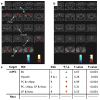
Effects of DBS on brain metabolism in the offspring of saline treated dams. Colored PET overlays on MR reference indicate increased 18F-FDG uptake (hot colors) or decreased (cold colors) glucose metabolism for DBS treated rats compared to sham rats. (a) mPFC-DBS in saline rats increased metabolic activity in the striatum (ST), ventral hippocampus (vHipp), parietal cortex (PC) and Nacc, and reduced it in the brainstem (BS), periaqueductal gray matter (PAG) hypothalamus (hypoth) and cerebellum (Cb). (b) Nacc-DBS in saline rats increased metabolic activity in the vHipp and olfactory bulb (OB), and decreased FDG-uptake in the septal area (SA), hypoth, BS and PAG. (c) Table summarizes statistical group differences for the respective regions of interest (ROI) and brain sides (right: R, left: L).
Fig. 5
Fig. 5
Effects of DBS on brain metabolism in the offspring poly I:C treated dams. Colored PET overlays on MR reference indicate increased 18F-FDG uptake (hot colors) or decreased (cold colors) glucose metabolism for DBS treated rats compared to sham rats. (a) mPFC-DBS in poly I:C rats increased metabolic activity in the straitum (ST), ventral hippocampus (vHipp), parietal cortex (PC) and Nacc, and decreased FDG-uptake in the brainstem (BS) and hypothalamus (hypoth). (b) Nacc-DBS in poly I:C rats did not induce any significant metabolic change in the brain. (c) Table summarizes statistical group differences for the respective regions of interest (ROI) and brain sides (right: R, left: L).

References

    1. Angelov SD, Dietrich C, Krauss JK, Schwabe K. Effect of deep brain stimulation in rats selectively bred for reduced prepulse inhibition. Brain Stimul. 2014;7:595–602. - PubMed
    1. Baharnoori M, Bhardwaj SK, Srivastava LK. Neonatal behavioral changes in rats with gestational exposure to lipopolysaccharide: a prenatal infection model for developmental neuropsychiatric disorders. Schizophr Bull. 2012;38:444–456. - PMC - PubMed
    1. Barak S, Weiner I. Putative cognitive enhancers in preclinical models related to schizophrenia: the search for an elusive target. Pharmacol Biochem Behav. 2011;99:164–189. - PubMed
    1. Ewing SG, Winter C. The ventral portion of the CA1 region of the hippocampus and the prefrontal cortex as candidate regions for neuromodulation in schizophrenia. Med Hypotheses. 2013;80:827–832. - PubMed
    1. Falkai P, Wobrock T, Lieberman J, Glenthoj B, Gattaz WF, Moller HJ. World federation of societies of biological psychiatry (WFSBP) guidelines for biological treatment of schizophrenia, part 1: acute treatment of schizophrenia. World J Biol Psychiatry. 2005;6:132–191. - PubMed

Publication types

MeSH terms