Review
. 2016 May 26:7:137.
doi: 10.3389/fphar.2016.00137. eCollection 2016.

Microneedles As a Delivery System for Gene Therapy

Affiliations
Review

Microneedles As a Delivery System for Gene Therapy

Wei Chen et al. Front Pharmacol. .

Abstract

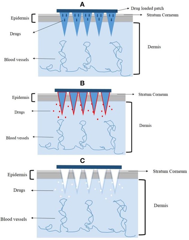
Gene delivery systems can be divided to two major types: vector-based (either viral vector or non-viral vector) and physical delivery technologies. Many physical carriers, such as electroporation, gene gun, ultrasound start to be proved to have the potential to enable gene therapy. A relatively new physical delivery technology for gene delivery consists of microneedles (MNs), which has been studied in many fields and for many molecule types and indications. Microneedles can penetrate the stratum corneum, which is the main barrier for drug delivery through the skin with ease of administration and without significant pain. Many different kinds of MNs, such as metal MNs, coated MNs, dissolving MNs have turned out to be promising in gene delivery. In this review, we discussed the potential as well as the challenges of utilizing MNs to deliver nucleic acids for gene therapy. We also proposed that a combination of MNs and other gene delivery approaches may lead to a better delivery system for gene therapy.

Keywords: approaches; delivery; gene; micronnedles; therapy.


Figures

Figure 1
Figure 1
Representative physical methods for transdermal delivery. (A) Ultrasound methods, (B) Intradermal injection, (C) Gene gun, (D) Microneedles.
Figure 2
Figure 2
The monolithic fabrication of a HEM by drawing lithography with antidromic isolation. (A) Liquid maltose was coated on a planar surface, and contacted with the 5 × 5 array electrodes of the HEMs as a drawing pillar. (B) The glassy maltose between Tm and Tg was elongated by drawing of electrodes. (C) After lowering the temperature to sub-Tg, the elongated 3D structures were cured to a solid state. (D) The coating surface was melted at a temperature greater than Tm to isolate elongated 3D structures from 2D coating surface, and this antidromic isolation fabricated dissolving microneedles of the HEMs. Adapted with permission from (Lee et al., 2011).
Figure 3
Figure 3
The mechanisms of different microneedles to deliver drugs. (A) The solid microneedles. (B) The coated microneedles. (C) The dissolving microneedles.

References

    1. Birchall J., Coulman S., Pearton M., Allender C., Brain K., Anstey A., et al. (2005). Cutaneous DNA delivery and gene expression in ex vivo human skin explants via wet-etch micro-fabricated micro-needles. J. Drug Target. 13, 415–421. 10.1080/10611860500383705 - DOI - PubMed
    1. Blaese R. M., Culver K. W., Miller A. D., Carter C. S., Fleisher T., Clerici M., et al. (1995). T lymphocyte-directed gene therapy for ADA- SCID: initial trial results after 4 years. Science 270, 475–480. 10.1126/science.270.5235.475 - DOI - PubMed
    1. Bryant L. M., Christopher D. M., Giles A. R., Hinderer C., Rodriguez J. L., Smith J. B., et al. (2013). Lessons learned from the clinical development and market authorization of Glybera. Hum. Gene Ther. Clin. Dev. 24, 55–64. 10.1089/humc.2013.087 - DOI - PMC - PubMed
    1. Castanotto D., Rossi J. J. (2009). The promises and pitfalls of RNA-interference-based therapeutics. Nature 457, 426–433. 10.1038/nature07758 - DOI - PMC - PubMed
    1. Cavalli R., Bisazza A., Lembo D. (2013). Micro- and nanobubbles: a versatile non-viral platform for gene delivery. Int. J. Pharm. 456, 437–445. 10.1016/j.ijpharm.2013.08.041 - DOI - PubMed