Review
. 2016 May 31:7:95.
doi: 10.3389/fgene.2016.00095. eCollection 2016.

Melanocortin 1 Receptor: Structure, Function, and Regulation

Affiliations
Review

Melanocortin 1 Receptor: Structure, Function, and Regulation

Erin M Wolf Horrell et al. Front Genet. .

Abstract

The melanocortin 1 receptor (MC1R) is a melanocytic Gs protein coupled receptor that regulates skin pigmentation, UV responses, and melanoma risk. It is a highly polymorphic gene, and loss of function correlates with a fair, UV-sensitive, and melanoma-prone phenotype due to defective epidermal melanization and sub-optimal DNA repair. MC1R signaling, achieved through adenylyl cyclase activation and generation of the second messenger cAMP, is hormonally controlled by the positive agonist melanocortin, the negative agonist agouti signaling protein, and the neutral antagonist β-defensin 3. Activation of cAMP signaling up-regulates melanin production and deposition in the epidermis which functions to limit UV penetration into the skin and enhances nucleotide excision repair (NER), the genomic stability pathway responsible for clearing UV photolesions from DNA to avoid mutagenesis. Herein we review MC1R structure and function and summarize our laboratory's findings on the molecular mechanisms by which MC1R signaling impacts NER.

Keywords: ASIP; ATR; DNA repair; MC1R; melanocortin; melanocyte; melanoma; βD3.


Figures

FIGURE 1
FIGURE 1
Melanocortin 1 receptor (MC1R) gene and protein structures. (A) The human MC1R locus (cytogenetic location: 16q24.3) encodes a seven transmembrane protein that is highly polymorphic. (B) The mature MC1R protein is a Gs-protein coupled receptor (GPCR) that spans the membrane seven times. Extracellular and transmembrane domains engage MC1R ligands while intracellular and transmembrane domains regulate adenylyl cyclase interactions and signaling.
FIGURE 2
FIGURE 2
Melanocortin – MC1R signaling axis. Melanocortins (α-MSH, ACTH) are produced basally by the pituitary and induced in the skin after UV injury. Binding of these melanocortin ligands to MC1R promotes critical UV-resistance physiologic changes in melanocytes and protects the skin from UV damage. Upon binding melanocortins, MC1R activates adenylyl cyclase and stimulates cAMP production. In turn, a variety of downstream effector pathways including induction of the CREB and Mitf transcription factor networks and an increase in the activity of PKA takes place. Expression of a variety of enzymes including tyrosinase (TYR) and dopachrome tautomerase (DCT) involved in melanin biosynthesis is increased and melanin production is up-regulated. Melanin produced in organelles termed melanosomes, is transferred to neighboring keratinocytes and in this way a UV-protective layer of pigment in the epidermis is established to enhance the skin’s ability to resist further UV injury. Importantly, melanocytic genomic stability is also enhanced through improved DNA repair. In the absence of a functional melanocortin signaling axis, these pathways are blunted, the skin is under-melanized and melanocytes accumulate more UV mutations as a result of ineffective DNA repair. In this way, individuals with inherited defects in MC1R signaling are at heightened risk for melanoma.
FIGURE 3
FIGURE 3
Melanin synthesis. There are two main types of melanin: the dark brown/black UV-protective eumelanin and the red/blonde sulfated pheomelanin pigment. Each is derived from progressive cyclization and oxidation of the amino acid tyrosine. Tyrosinase, the rate-limiting enzyme for melanogenesis, catalyzes the first two stages of melanin biosynthesis. When MC1R is functional and cAMP levels are high, melanocytes produce eumelanin preferentially. In contrast, when MC1R is dysfunctional and cAMP levels are low, cysteine is incorporated and pheomelanin is made instead. Important pigment enzymes which cause hypopigmentary phenotypes when defective include tyrosinase (TYR), dopachrome tautomerase (DCT), and tyrosinase related protein-1 (TRP1). Complexion and UV resistance are mainly determined by the amount of eumelanin in the skin.
FIGURE 4
FIGURE 4
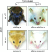
Melanocortin 1 receptor controls coat color in C57BL/6 mice. C57BL/6 mice with wild type (functional) Mc1r exhibit a eumelanotic phenotype, whereas congenic animals defective for Mc1r, such as extension mice, exhibit a pheomelanotic coat color. Shown are Mc1rE/E (wt) and Mc1re/e (extension) age-matched animals along with C57BL/6 Tyrc2j/c2j animals in either the Mc1rE/E (wt) or Mc1re/e (extension) background in which neither eumelanin nor pheomelanin can be made because of defective tyrosinase function (Jarrett et al., 2014). Note that these animals express the K14-Scf transgene resulting in retention of interfollicular epidermal melanocytes and pigmentation of the skin in addition to the fur.
FIGURE 5
FIGURE 5
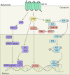
Melanocortin 1 receptor signaling and melanocyte genomic integrity. MC1R signaling promotes genomic stability through multiple mechanisms. MC1R activation induces translocation of NR4A2 to the nucleus in a p38 and PARP1 dependent manner where it co-localizes with XPC and XPE at sites of UV induced DNA damage. MC1R activation also leads to elevated levels of XPC and γH2Ax promoting the formation of DNA repair complexes. Levels of γH2AX are regulated by Wip1 downstream of ATR and DNA-PK mediated phosphorylation of p53 at S15. In addition, PKA activation promotes the phosphorylation of ATR at S435. pS435-ATR complexes with XPA in the nucleus. Following phosphorylation of XPA, the complex translocates to sites of UV induced DNA damage to accelerate and enhance nucleotide excision repair (NER).
FIGURE 6
FIGURE 6
Melanocortin 1 receptor ligand interactions. Three major MC1R ligands are α-MSH, ASIP, and βD3. The melanocortin α-MSH functions as a positive agonist to increase cAMP levels downstream of MC1R. ASIP is a potent negative agonist that decreases basal MC1R signaling and interferes with melanocortin-induced cAMP upregulation. βD3 is thought to act as a neutral antagonist of MC1R, blocking the interactions with either melanocortins or ASIP but having little signaling impact independently. Each ligand appears to function as a competitive inhibitor for the others with only one able to bind to MC1R at any given time. Please note that although not shown in this diagram, ACTH’s ability to agonize MC1R is roughly the same as that of α-MSH.
FIGURE 7
FIGURE 7
Melanocortin synthesis through proopiomelanocortin (POMC) processing. Melanocortins are derived from the proopiomelanocortin (POMC) precursor peptide that is cleaved into functional peptide fragments by a pair of serine protease pro-protein convertases, PC1 and PC2. PC1 cleaves POMC into four subunits including an n-terminal region from which γ-MSH is derived, adrenocorticotropic hormone (ACTH) and β-lipotropin (β-LPH) from which γ-LPH, β-MSH and β-endorphin are generated by PC2 cleavage. α-MSH represents the first 13 amino acids of ACTH and is dependent on PC2 activity for its synthesis.

References

    1. Abdel-Malek Z., Scott M. C., Suzuki I., Tada A., Im S., Lamoreux L., et al. (2000). The melanocortin-1 receptor is a key regulator of human cutaneous pigmentation. Pigment Cell Res. 13(Suppl. 8), 156–162. 10.1034/j.1600-0749.13.s8.28.x - DOI - PubMed
    1. Abdel-Malek Z. A., Kadekaro A. L., Kavanagh R. J., Todorovic A., Koikov L. N., McNulty J. C., et al. (2006). Melanoma prevention strategy based on using tetrapeptide alpha-MSH analogs that protect human melanocytes from UV-induced DNA damage and cytotoxicity. FASEB J. 20 1561–1563. 10.1096/fj.05-5655fje - DOI - PubMed
    1. Abdel-Malek Z. A., Ruwe A., Kavanagh-Starner R., Kadekaro A. L., Swope V., Haskell-Luevano C., et al. (2009). alpha-MSH tripeptide analogs activate the melanocortin 1 receptor and reduce UV-induced DNA damage in human melanocytes. Pigment Cell Melanoma Res. 22 635–644. 10.1111/j.1755-148X.2009.00598.x - DOI - PubMed
    1. Abdel-Malek Z. A., Scott M. C., Furumura M., Lamoreux M. L., Ollmann M., Barsh G. S., et al. (2001). The melanocortin 1 receptor is the principal mediator of the effects of agouti signaling protein on mammalian melanocytes. J. Cell Sci. 114 1019–1024. - PubMed
    1. Abdel-Malek Z. A., Swope V. B., Starner R. J., Koikov L., Cassidy P., Leachman S. (2014). Melanocortins and the melanocortin 1 receptor, moving translationally towards melanoma prevention. Arch. Biochem. Biophys. 563 4–12. 10.1016/j.abb.2014.07.002 - DOI - PubMed