

Case Reports
doi: 10.2484/rcr.v2i4.119.
eCollection 2007.
Epithelioid Hemangioendothelioma of the Distal Radius: A Case Report
- PMID: 27303497
- PMCID: PMC4895778
- DOI: 10.2484/rcr.v2i4.119
Item in Clipboard
Case Reports
Epithelioid Hemangioendothelioma of the Distal Radius: A Case Report
Radiol Case Rep.
.
Abstract
Epithelioid hemangioendothelioma is a rare vascular tumor with cytologic behavior between angiosarcoma and hemangioma. We present the case of a 58-year-old male with primary epithelioid hemangioendothelioma of the distal radius measuring 6.2 × 5 cm with extension into the pronator quadratus and brachioradialis muscles. We discuss our approach to performing a limb-sparing resection combined with reconstruction to preserve upper extremity function. A review of the clinical, radiographic, and pathologic features of epithelioid hemangioendothelioma is also presented.
Keywords: CT, computed tomography; EH, epithelioid hemangioendothelioma; MRI, magnetic resonance imaging.
Figures
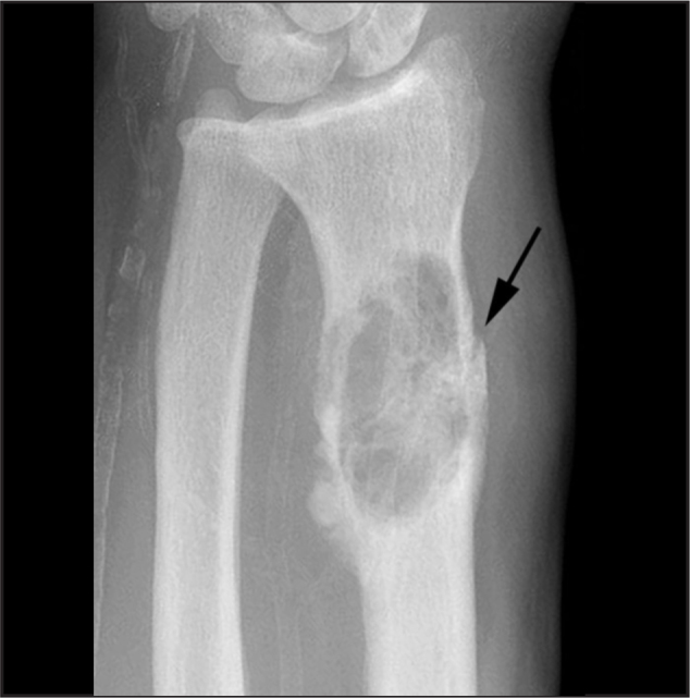
Preoperative oblique radiograph of the right distal forearm demonstrating an epithelioid hemangioendothelioma. A lytic, septated, expansile lesion is present without identifiable matrix. A pathologic fracture is visible along the radial border.
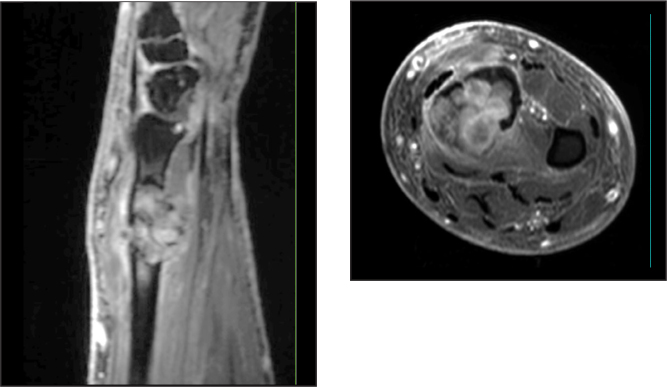
Sagittal (A) and axial (B) T1-weighted fat suppressed MR images with gadolinium enhancement demonstrate a heterogeneously enhancing, aggressive lesion that traverses the bone cortex and is encased by the pronator quadratus and brachioradialis muscles.
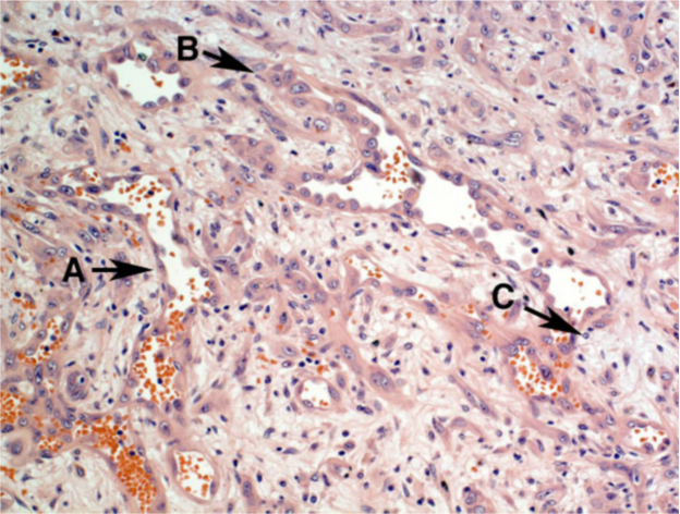
Hematoxylin and eosin stain of the high grade epithelioid hemangioendothelioma. Note is made of epithelioid neoplastic cells with prominent nuclei and nucleoli lining the vascular spaces, with eosinophilic and amphophilic cytoplasms. Focal myxoid or chondroid-like matrix is present. A: Vasoformative channels. B: Cytologic atypia, the nuclei are plump, hyperchromatic, variable in shape, and may bulge into the vascular lumina. C: Collagenized, chondroid appearing stroma.
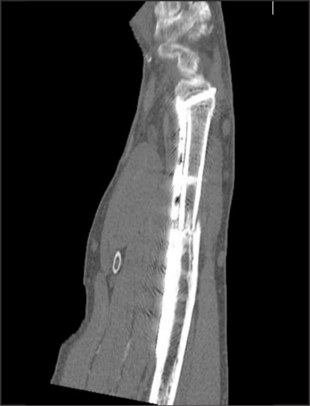
CT scan obtained 9 months postoperatively. The distal aspect of the graft is well incorporated. The proximal aspect demonstrates partial incorporation with ingrowth into the medullary canal and incomplete callus formation at the periphery. Heterotopic ossification is seen adjacent to the plate.
References
-
- Enzinger F. Hemangioendothelioma: vascular tumors of intermediate malignancy. 3rd ed. Mosby; St Louis: 1995.
-
- Evans HL, Raymond AK, Ayala AG. Vascular tumors of bone: A study of 17 cases other than ordinary hemangioma, with an evaluation of the relationship of hemangioendothelioma of bone to epithelioid hemangioma, epithelioid hemangioendothelioma, and high-grade angiosarcoma. Hum Pathol. 2003;34(7):680–689. [PubMed] - PubMed
Publication types
LinkOut - more resources
Full Text Sources
