Case Reports
. 2015 Dec 7;3(2):153.
doi: 10.2484/rcr.v3i2.153. eCollection 2008.

Coincident Giant Cavernous Angioma and Large Middle Cerebral Artery Aneurysm

Case Reports

Coincident Giant Cavernous Angioma and Large Middle Cerebral Artery Aneurysm

Tom D Newton et al. Radiol Case Rep. .

Abstract

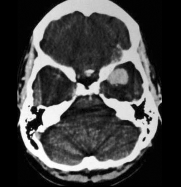
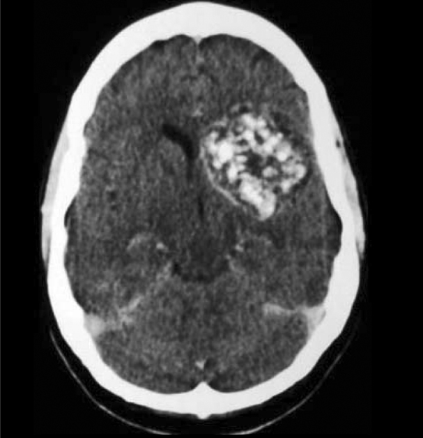
Cavernous angiomas although relatively common lesions rarely reach a large size. They have a well documented association with AVMs, capillary telangiectases and venous angiomas but are not specifically associated with intracerebral aneurysms. We present a case of what we believe to be the 4th largest reported giant cavernous angioma to present in adulthood. This cavernous angioma also happened to be associated with a large intracerebral aneurysm, an association not previously reported. The sometimes confusing nomenclature of cavernous angiomas and other similar vascular malformations is also discussed.

Keywords: AVM, arteriovenous malformation; CT, computed tomography; MRI, magnetic resonance imaging.


Figures

Figure 1A
Figure 1A
43-year-old woman with worsening left-sided headaches. Post-contrast CT scan shows left MCA aneurysm.
Figure 1B
Figure 1B
43-year-old woman with worsening left-sided headaches. Post-contrast CT scan shows left cavernous angioma.
Figure 2A
Figure 2A
43-year-old woman with worsening left-sided headaches. Axial T2W images demonstrate MCA aneurysm which was originally mistaken for a “tumour nodule.”
Figure 2B
Figure 2B
43-year-old woman with worsening left-sided headaches. Axial T2W images demonstrate typical features of a cavernous angioma with a mixed signal core, complete haemosiderin rim, and blooming susceptibility effect.
Figure 3A
Figure 3A
43-year-old woman with worsening left-sided headaches. Axial T1W pre-contrast (gradient echo) MRI. Note the marked heterogeneity within the cavernous angioma, the high signal change which is consistent with blood products.
Figure 3B
Figure 3B
43-year-old woman with worsening left-sided headaches. Axial T1W post-contrast (spin-echo) MRI. No contrast enhancement is demonstrated.
Figure 4
Figure 4
43-year-old woman with worsening left-sided headaches. T1W post-contrast coronal MRI clearly demonstrating the non-enhancing cavernoma and the immediately subjacent enhancing aneurysm.
Figure 5
Figure 5
43-year-old woman with worsening left-sided headaches. Selective left internal carotid artery angiogram clearly depicting the large left middle cerebral artery aneurysm. The cavernous angioma is angiographically occult.

References

    1. Otten P, Pizzolato GP, Rilliet B, Berney J. 131 cases of cavernous angioma of the CNS, discovered by retrospective analysis of 24,535 autopsies. Neurochirurgie. 1989;35(2):82–83. [PubMed] 128-31. - PubMed
    1. Zabramski JM, Wascher TM, Spetzler RF, Johnson B, Golfinos J, Drayer BP. The natural history of familial cavernous malformations: results of an ongoing study. J Neurosurg. 1994;80(3):422–432. [PubMed] - PubMed
    1. medcyclopedia.com. Venous and cavernous angiomas – Imaging. [Accessed 27 May 2008]
    1. Osborn AG, Blaser SI, Salzman KL. Pocket radiologist. Brain: Top 100 diagnoses. WB Saunders; Philadelphia: 2002.
    1. Del Curling O, Jr., Kelly DL, Jr., Elster AD, Craven TE. An analysis of the natural history of cavernous angiomas. J Neurosurg. 1991;75(5):702–708. [PubMed] - PubMed

Publication types

LinkOut - more resources