

Case Reports
doi: 10.2484/rcr.v3i2.164.
eCollection 2008.
Lipoma Arborescens of the Knee in a 17-Year-Old Man
- PMID: 27303521
- PMCID: PMC4896232
- DOI: 10.2484/rcr.v3i2.164
Item in Clipboard
Case Reports
Lipoma Arborescens of the Knee in a 17-Year-Old Man
Radiol Case Rep.
.
Abstract
We present a case of lipoma arborescens of the knee in a 17-year-old man, discuss its characteristic imaging findings, and review the relevant differential diagnoses.
Keywords: MRI, magnetic resonance imaging; PVNS, pigmented villonodular synovitis; STIR, short tau inversion recovery.
Figures

Lipoma arborescens in a 17-year-old man. Lateral radiograph of the knee shows a large mass within the suprapatellar region with areas of soft tissue and fat density.

Lipoma arborescens in a 17-year-old man. Sagittal T1 weighted MRI shows a large suprapatellar effusion with a frond-like synovial mass of fat intensity.

Lipoma arborescens in a 17-year-old man. Sagittal proton density MRI shows the large high-signal effusion with synovial proliferation.

Lipoma arborescens in a 17-year-old man. Sagittal STIR MRI shows the synovial mass to be the same low intensity as fat.
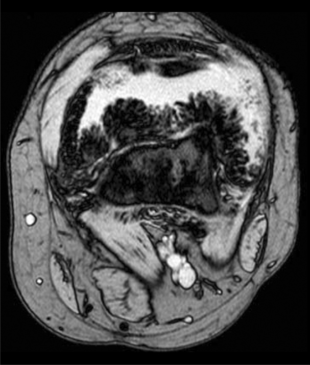
Lipoma arborescens in a 17-year-old man. Axial gradient echo MRI shows the characteristic frond-like pattern.

Lipoma arborescens in a 17-year-old man. Coronal T1 fat-suppression MRI following gadolinium injection shows no enhancement of the synovial mass but enhancement of the effusion.

Lipoma arborescens in a 17-year-old man. Sagittal T1 fat-suppression MRI following gadolinium injection shows no enhancement of the synovial mass but enhancement of the effusion.
References
Publication types
LinkOut - more resources
Full Text Sources
