Case Reports
. 2015 Nov 6;3(3):183.
doi: 10.2484/rcr.v3i3.183. eCollection 2008.

Reactivation Mycobacterium Tuberculosis Presenting as Empyema Necessitans 55 Years Following Thoracoplasty

Case Reports

Reactivation Mycobacterium Tuberculosis Presenting as Empyema Necessitans 55 Years Following Thoracoplasty

Nancy J McNulty. Radiol Case Rep. .

Abstract

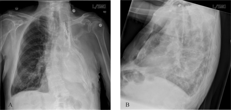
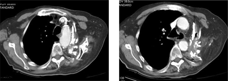
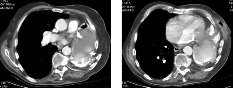
We describe the case of a 79-year-old man who presented with an enlarging mass on his chest wall. He had a history of thoracoplasty performed 55 years ago for treatment of pulmonary tuberculosis. The mass was subsequently proven to be the result of empyema neccesitans caused by reactivation tuberculosis. Empyema neccesitans is a well described entity in which an empyema spontaneously decompresses by dissecting into the chest wall and extrathoracic soft tissues. This can occur following necrotizing pneumonia, including pyogenic or tuberculus, or pulmonary abscess. Complications from collapse therapy for tuberculosis can be encountered decades following the surgery, however, empyema necessitans due to reactivation tuberculosis is rare. This case affords the opportunity to review the goals, techniques, and radiologic appearance of thoracoplasty.

Keywords: CT, computed tomography; TB, tuberculosis.


Figures

Figure 1A-B
Figure 1A-B
79-year-old man with reactivation tuberculosis and empyema necessitans 55 years following thoracoplasty. PA and lateral CXR demonstrate left upper rib resections, collapse of the left chest wall, opacification and volume loss of the left hemithorax and chest wall calcifications.
Figures 2A-B
Figures 2A-B
79-year-old man with reactivation tuberculosis and empyema necessitans 55 years following thoracoplasty. Axial CT images of the chest show resections of left upper ribs, distortion and collapse of the left upper thorax and apposition of the chest wall to the superior mediastinum.
Figures 2C-D
Figures 2C-D
79-year-old man with reactivation tuberculosis and empyema necessitans 55 years following thoracoplasty. Axial CT images of the chest show the hemithorax opacified with fluid, peripheral lobular areas of high attenuation tissue, and calcification. A subcutaneous abscess cavity is present.

References

    1. Chaiyasate K, Hramiec J. Images in Clinical Medicine. Tuberculosis empyema necessitatis. NEJM. 2005 March 3;352(9):e8. [PubMed] - PubMed
    1. Dewan RK, Singh S, Kumar A, Meena BK. Thoracoplasty: An Obsolete Procedure? The Indian Journal of Chest Diseases & Allied Sciences. 1999 April-June;41(2):83–88. [PubMed] - PubMed
    1. Hopkins RA, Ungerleider RM, Staub EW, Yound WG. The Modern Use of Thoracoplasty. The Annals of thoracic surgery. 1985 Aug;40(2):181–187. [PubMed] - PubMed
    1. Barker WL. Thoracoplasty. Chest Surgery Clinics of North America, August. 1994;4(3):593–615. [PubMed] - PubMed
    1. Pomerantz M, Mault JR. History of Resectional Surgery for Tuberculosis and Other Mycobacterial Infections. Chest Surgery Clinics of North America, February. 2000;10(1):131–133. [PubMed] - PubMed

Uncited Reference

    1. Weissberg D, Weissberg D. Late Complications of Collapse Therapy for Pulmonary Tuberculosis. Chest. 2001 Sep;120(3):696–697. [PubMed] - PubMed

Publication types

LinkOut - more resources