

Case Reports
doi: 10.2484/rcr.v3i3.195.
eCollection 2008.
Fibrolipomatous Hamartoma of the Median Nerve
- PMID: 27303544
- PMCID: PMC4897024
- DOI: 10.2484/rcr.v3i3.195
Item in Clipboard
Case Reports
Fibrolipomatous Hamartoma of the Median Nerve
Radiol Case Rep.
.
Abstract
We present the case of a 33-year-old woman who presented with a slowly enlarging mass over the volar aspect of the wrist that had been present since infancy which for the previous year had been causing progressive pain. The lesion was proven to be a fibrolipomatous hamartoma, a rare benign tumor that most commonly affects the median nerve. We discuss the characteristic radiologic appearance of this entity that is often pathognomonic and allows a confident diagnosis without the need for biopsy.
Keywords: CT, computed tomography; MRI, magnetic resonance imaging.
Figures
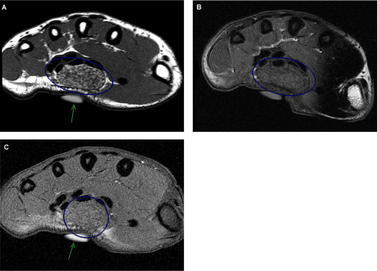
33-year-old woman with fibrolipomatous hamartoma of the median nerve. Axial T1 weighted pre (A) and post (C) gadolinium (TR 400/TE 13) and T1 fat-suppressed (C) (367/12) fast spin echo (FSE) MRI at the level of the carpal tunnel in location of palpable mass (vitamin E capsule denoted with green arrow). There is a fusiform mass (blue circle) in the expected location of the median nerve that is continuous with the nerve proximally and distally. On the non fat-suppressed images the high signal represents the fatty tissue infiltrating and enlarging the median nerve which drops completely with fat suppression. The fat is interspersed within thickened nerve fascicles which are low intensity. The mass displaces the flexor tendons.
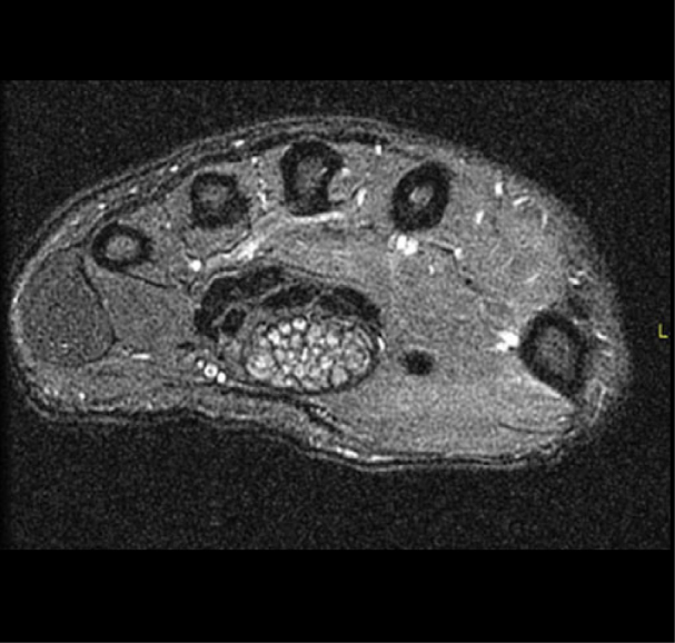
33-year-old woman with fibrolipomatous hamartoma of the median nerve. Axial T2 weighted (3517/49) fat suppressed FSE MRI shows intermediate signal longitudinally oriented cylindrical structures representing the nerve fascicles. There is fusiform enlargement of nerve bundles caused by fatty infiltration.

33-year-old woman with fibrolipomatous hamartoma of the median nerve. Coronal T1 (500/11) FSE MRI shows thickened nerve fascicles with high signal fat interspersed (blue circle). The mass displaces the flexor tendons.

33-year-old woman with fibrolipomatous hamartoma of the median nerve. Sagittal T1 (600/11) FSE MRI shows mass (blue arrow) displacing the flexor retinaculum anteriorly.
References
Publication types
LinkOut - more resources
Full Text Sources
