. 2015 Dec 2;1(1):e00028-15.
doi: 10.1128/mSphere.00028-15. eCollection 2016 Jan-Feb.

Novel Immunomodulatory Flagellin-Like Protein FlaC in Campylobacter jejuni and Other Campylobacterales

Affiliations

Novel Immunomodulatory Flagellin-Like Protein FlaC in Campylobacter jejuni and Other Campylobacterales

Eugenia Faber et al. mSphere. .

Abstract

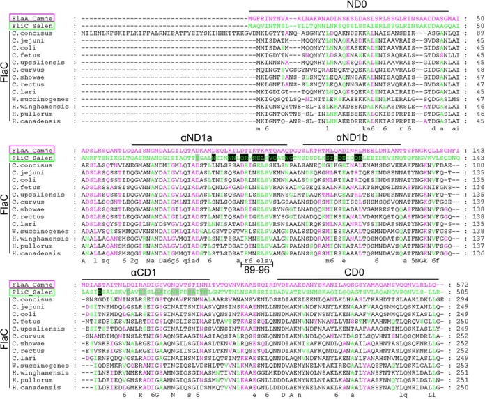
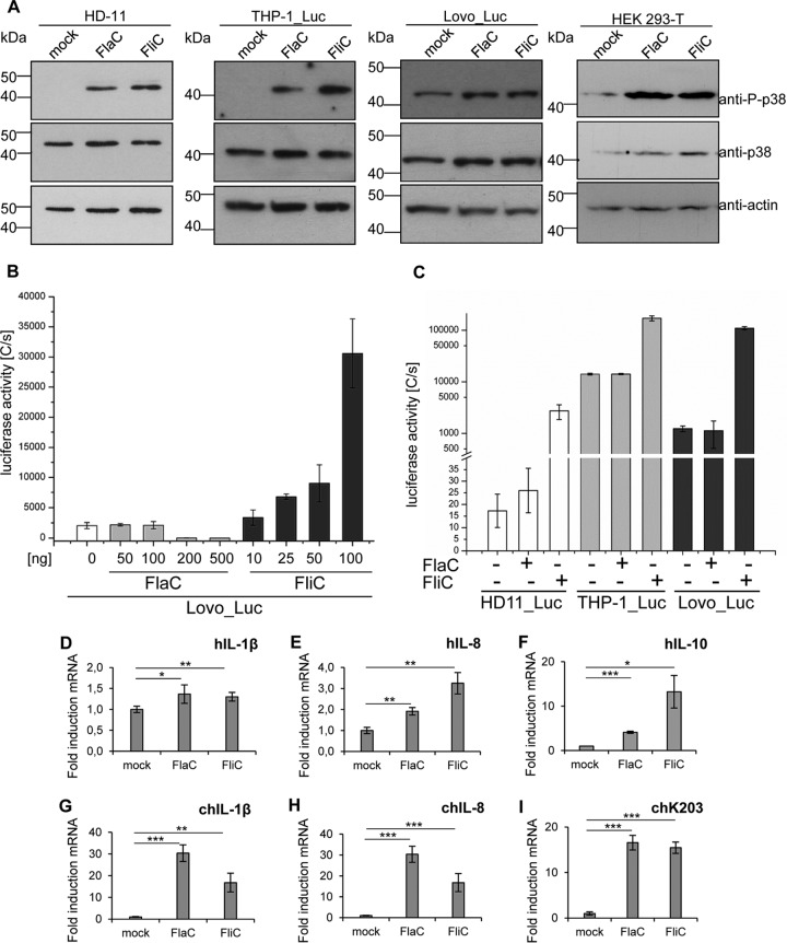
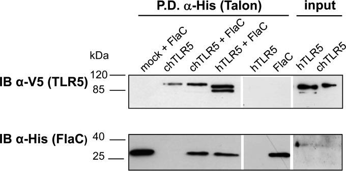
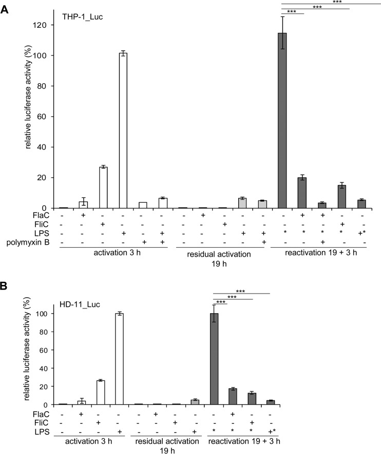
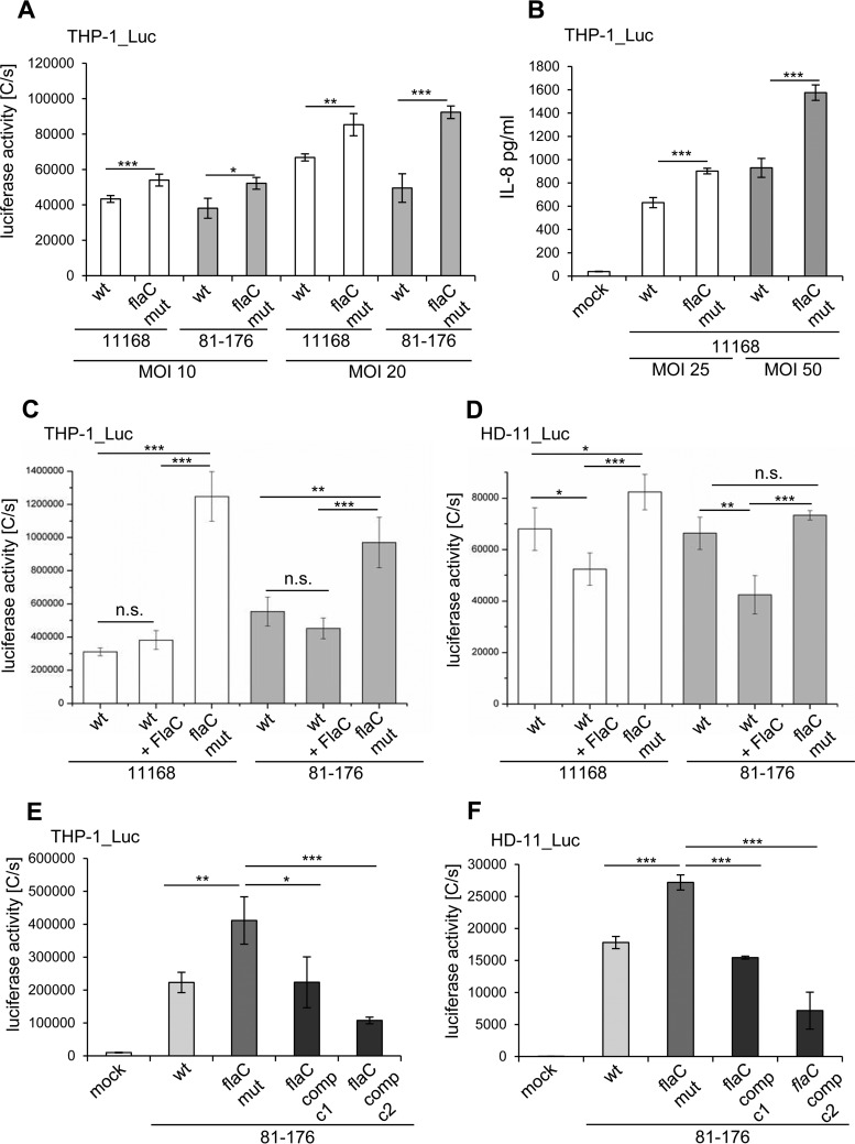
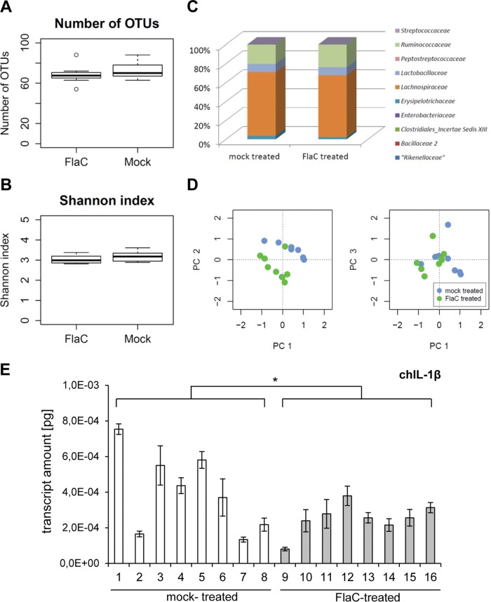
The human diarrheal pathogens Campylobacter jejuni and Campylobacter coli interfere with host innate immune signaling by different means, and their flagellins, FlaA and FlaB, have a low intrinsic property to activate the innate immune receptor Toll-like receptor 5 (TLR5). We have investigated here the hypothesis that the unusual secreted, flagellin-like molecule FlaC present in C. jejuni, C. coli, and other Campylobacterales might activate cells via TLR5 and interact with TLR5. FlaC shows striking sequence identity in its D1 domains to TLR5-activating flagellins of other bacteria, such as Salmonella, but not to nonstimulating Campylobacter flagellins. We overexpressed and purified FlaC and tested its immunostimulatory properties on cells of human and chicken origin. Treatment of cells with highly purified FlaC resulted in p38 activation. FlaC directly interacted with TLR5. Preincubation with FlaC decreased the responsiveness of chicken and human macrophage-like cells toward the bacterial TLR4 agonist lipopolysaccharide (LPS), suggesting that FlaC mediates cross-tolerance. C. jejuni flaC mutants induced an increase of cell responses in comparison to those of the wild type, which was suppressed by genetic complementation. Supplementing excess purified FlaC likewise reduced the cellular response to C. jejuni. In vivo, the administration of ultrapure FlaC led to a decrease in cecal interleukin 1β (IL-1β) expression and a significant change of the cecal microbiota in chickens. We propose that Campylobacter spp. have evolved a novel type of secreted immunostimulatory flagellin-like effector in order to specifically modulate host responses, for example toward other pattern recognition receptor (PRR) ligands, such as LPS. IMPORTANCE Flagellins not only are important for bacterial motility but are major bacterial proteins that can modulate host responses via Toll-like receptor 5 (TLR5) or other pattern recognition receptors. Campylobacterales colonizing the intestinal tracts of different host species harbor a gene coding for an unusual flagellin, FlaC, that is not involved in motility but is secreted and possesses a chimeric amino acid sequence composed of TLR5-activating and non-TLR5-activating flagellin sequences. Campylobacter jejuni FlaC activates cells to increase in cytokine expression in chicken and human cells, promotes cross-tolerance to TLR4 ligands, and alters chicken cecal microbiota. We propose that FlaC is a secreted effector flagellin that has specifically evolved to modulate the immune response in the intestinal tract in the presence of the resident microbiota and may contribute to bacterial persistence. The results also strengthen the role of the flagellar type III apparatus as a functional secretion system for bacterial effector proteins.

Keywords: Campylobacter; TLR5; flagellin; host-pathogen interaction; immune response.


Figures

FIG 1
FIG 1
ClustalOmega alignment of FlaC protein sequences. C. jejuni (Camje) FlaA (pink, a representative of non-TLR5-stimulatory flagellins), S. enterica (Salen) FliC (green, a paradigm for a TLR5-activating flagellin), and FlaC protein sequences of various Epsilonproteobacteria were aligned with ClustalOmega (http://www.ebi.ac.uk/Tools/msa/clustalo) and visualized in GeneDoc (https://www.psc.edu/index.php/user-resources/software/genedoc). The locations of the D0 and D1 domains are indicated above the sequences. Residues of FliC involved in TLR5 binding and activation are shaded in gray (primary interface A) and in black (primary interface B) (according to reference 12). Residues of FlaC identical to those in C. jejuni FlaA or S. enterica FliC are colored accordingly in pink or green, respectively. A consensus score is shown underneath the alignment. Only flagellin sequence domains D0 and D1 present in FlaC are depicted; since the D2 and D3 domains are largely absent from FlaC orthologues, these domains have been omitted from the alignment.
FIG 2
FIG 2
Characterization of C. jejuni flaC mutants and subcellular localization of FlaC. (A) Motility of the C. jejuni 11168 and 81-176 wild types (wt) and two corresponding flaC mutants (clone 1 and 2 [c1 and c2, respectively]) of each strain. A representative soft-agar motility plate from at least three independent assays shows the motility areas of all bacterial strains (swim diameter of ca. 10 mm) after 2 days of incubation at 37°C (scale bar, 10 mm). Corresponding motility-negative controls of C. jejuni (flgR mutant) always exhibited a swim halo diameter of <1 mm under the same conditions, while flaC-complemented bacteria reproducibly had a swim diameter similar to that of the wild type and flaC mutant (not shown). (B to D) Subcellular localization of FlaC in C. jejuni bacterial fractions. Comparative Western blot analyses of whole-cell lysates and different fractions of C. jejuni 11168 and 81-176 wild-type and flaC-mutants grown under microaerobic conditions were performed using polyclonal rabbit FlaC antiserum (dilution, 1:5,000). (B) S, soluble bacterial fraction; IS, insoluble bacterial fraction. (C) Surface/flagellar proteins. (D) Secreted proteins. Expression of FlaC was also investigated under anaerobic conditions. Western blot analyses of whole-cell lysates and different fractions of wild-type and flaC mutants grown under anaerobic conditions yielded comparable results (not shown). Our estimate from comparative Western blots was that approximately 2 µg of FlaC was secreted per 109 bacteria, and 100 to 200 ng of cell-bound FlaC was present in the same number of bacteria.
FIG 3
FIG 3
Immune response in infected chickens to C. jejuni FlaC. (A) Adaptive immune response to recombinant C. jejuni FlaC in Campylobacter-infected chickens. Western blot analyses were performed to investigate and compare the reactivities of whole sera from Campylobacter-infected (lanes 1 to 19) and noninfected (lanes 20 to 22) chickens. Chicken sera were used at a dilution of 1:10,000 and detected with horseradish-peroxidase-coupled anti-chicken antibody. (B) In vivo expression of flaC in chicken cecal tissue. Transcript levels of flaC were determined by using quantitative RT-PCR of total RNA extracted from the ceca of five chickens which had been experimentally infected with C. jejuni strain RB922 (48). The amounts of specific flaC cDNA (in picograms) were normalized to C. jejuni 16S rRNA gene amounts determined in each animal.
FIG 4
FIG 4
Effect of purified C. jejuni FlaC on human and chicken cell signal transduction. (A) p38 MAP kinase phosphorylation in human Lovo_Luc, THP-1_Luc, HEK293T, and chicken HD-11 cells was analyzed by Western immunoblotting after coincubation of the cell lines with ultrapure recombinant Salmonella FliC (100 ng) or ultrapure recombinant C. jejuni FlaC (200 ng) for 4 h (see Materials and Methods). Densitometry of P-p38 band intensities was performed, and results were normalized against those for the respective p38 signal and the actin loading control band in each lane. Normalized intensity values indicated that for all 3 cell lines, P-p38 was at least 5-fold enhanced by FlaC over levels in the mock-incubated control. (B) Analysis of the concentration-dependent response of Lovo cells, stably transfected with an NF-κB luciferase reporter gene, toward Salmonella FliC (positive control) or recombinant C. jejuni FlaC. (C) HD-11, THP-1, and Lovo cells stably transfected with an NF-κB luciferase reporter were coincubated with Salmonella FliC (25 ng) or recombinant C. jejuni FlaC (100 ng) for 3 h in 96-well plates and analyzed for NF-κB-driven luciferase expression using the SteadyGlo luciferase assay. The luciferase activities in panels B and C are given in luminescence values as photon counts per second (C/s). (D to I) Quantitative RT-PCR of cytokine mRNA induction by C. jejuni FlaC in human and chicken cells. Levels of induction of hIL-1β (D), hIL-8 (E), and hIL-10 (F) by C. jejuni FlaC and S. Typhimurium FliC in human macrophages (THP-1) and of chIL-1β (G), chIL-8 (H), and chK203 (I) in chicken macrophages (HD-11) are shown. Both cell types were stimulated for 2 h with recombinant ultrapure proteins FlaC (500 ng) and FliC (300 ng). Isolated RNA was analyzed by quantitative RT-PCR. Transcript values were normalized to human or chicken GAPDH values and are presented as fold increases of mRNA levels compared to the level in a mock-coincubated control. Mean values and standard deviations from triplicate measurements are shown. Significant P values are indicated by asterisks (Student’s t test, unpaired, one-sided) as follows: *, 0.01 ≤ P ≤ 0.05; **, 0.001 ≤ P ≤ 0.01; and ***, P ≤ 0.001.
FIG 5
FIG 5
Interaction of C. jejuni FlaC with TLR5 in vitro. Binding of C. jejuni FlaC to TLR5 was tested by pulldown assay. Cleared cell lysates from TLR5 plasmid-transfected (hTLR5, human; chTLR5, chicken; both expressed as a V5-tagged protein fusion) or empty-plasmid-transfected cells were generated and coincubated with purified C. jejuni 6×His-FlaC. Pulldown against the 6×His tag fused to FlaC was performed using Talon Co2+ resin. Protein detection on Western blots was performed using rabbit anti-6×His antiserum (diluted 1:1,000; Rockland) for FlaC, and anti-V5 antibody (mouse monoclonal, 1:5,000; Invitrogen) for the detection of hTLR5 and chTLR5 (V5 tagged). The input was cleared cell lysates of TLR5-V5 plasmid-transfected HEK293T cells that were used for the pulldown. IB, immunoblotting; P.D., pulldown.
FIG 6
FIG 6
C. jejuni FlaC antagonizes the activating effect of the TLR4 ligand. Human (THP-1) (A) and chicken (HD-11) (B) macrophages stably transfected with the NF-κB luciferase reporter gene were coincubated with recombinant C. jejuni FlaC (100 ng), Salmonella FliC (50 ng), or E. coli LPS (25 ng) for 3 h, and NF-κB activation was measured in a SteadyGlo luciferase assay. Residual activation of the cells in all wells was determined by a luciferase measurement 19 h after the initial incubation. To analyze the reactivation potential, cells were preincubated with recombinant C. jejuni FlaC, Salmonella FliC, or E. coli LPS for 19 h and then coincubated with E. coli TLR4 ligand LPS (25 ng). The resulting reactivation potential of NF-κB was measured 3 h after the secondary coincubation step. As a control for TLR4-specific activity, control wells were preincubated with polymyxin B (10 µg/ml) 1 h before the initial activation, as indicated below the graph. For all measurements, relative luciferase activity is depicted as the percentage of maximal activation by LPS, which was defined as 100%. +, addition of substance on day 1; −, no addition of substance on day 1; *, addition of E. coli LPS on day 2. Significant P values are indicated by asterisks (Student’s t test, unpaired, two-sided), as follows: ***, P ≤ 0.001.
FIG 7
FIG 7
C. jejuni flaC mutants induce increased cell activation in human and chicken cells. (A and B) Comparison of cell activation of live C. jejuni with the respective flaC mutants of two strains, 11168 and 81-176. (A) Human THP-1 NF-κB luciferase reporter cells were coincubated with live C. jejuni bacteria of strain 11168 or 81-176 (wt) and corresponding isogenic flaC mutants (flaC mut) at different multiplicities of infection (MOI) for 3 h. NF-κB activation was determined using SteadyGlo luciferase substrate. Means and standard deviations from technical quadruplicates are shown as luciferase activity in counts per second (C/s). (B) IL-8 secretion induced by the live C. jejuni wild type and flaC mutants was measured by IL-8 ELISA. The statistical significance of differences was determined using Student’s t test (unpaired, one-sided). ***, P ≤ 0.001; **, 0.001 ≤ P ≤ 0.01; *, 0.01 ≤ P ≤ 0.05. (C and D) Human (THP-1) (C) and chicken (HD-11) (D) NF-κB luciferase reporter cell lines were coincubated with cleared lysate fractions of wild-type bacteria (wt) and corresponding flaC mutants (flaC mut) (100 ng of cleared lysate) of two different C. jejuni strains (11168, 81-176) for 3 h. For comparison, cells which were preincubated for 1 h with purified FlaC (100 ng) were also incubated with soluble fractions of wild-type lysates (100 ng) for 3 h. Luciferase activities were measured in a SteadyGlo luciferase assay. Mean values and standard deviations of triplicate measurements are depicted as luciferase activities (counts per second [C/s]). n.s., not significant. (E and F) Complementation of flaC restores the cell activation level by C. jejuni. (E) Human (THP-1) and (F) chicken (HD-11) macrophages stably transfected for the NF-κB luciferase reporter gene were coincubated for 3 h with soluble lysate fractions (100 ng) of the C. jejuni parental strain (wt), corresponding flaC mutant (flaC mut), and two flaC complementation clones of strain 81-176 (flaC comp, c1, and c2). NF-κB activation was measured using SteadyGlo substrate. Means and standard deviations from technical quadruplicates are shown as luciferase activity in counts per second [C/s]. Statistical significance was determined using Student’s t test (unpaired, two-sided). Significant P values are indicated by asterisks, as follows: *, 0.01 ≤ P ≤ 0.05; **, 0.001 ≤ P ≤ 0.01; ***, P ≤ 0.001.
FIG 8
FIG 8
FlaC has a significant influence on the chicken cecal microbiota and cecal expression of chicken IL-1β. Microbiota analysis of chicken cecal tissue was performed using 16S rRNA gene amplicon sequencing. The microbiotas were compared between a chicken group (8 animals) of FlaC-treated animals and 8 animals that were mock (PBS) treated. Shown are comparative numbers of OTU (A), a comparison of the within-sample OTU diversity between the two groups (Shannon index) (B), and the comparative family assignments of OTU present in the cecal microbiotas of the two groups (C). (D) A principal-coordinates analysis (PC; based on Bray-Curtis distances [see Text S1 in the supplemental material]) of the microbiota data indicated that the chickens in the FlaC-treated group (neon-green dots) have a significantly different microbiota composition than the chickens in the mock-treated group (light-blue dots). Depicted in the graphs are the distances between the microbiota data in the first three dimensions (axes PC 1 to PC 3) of the Bray-Curtis principal-coordinates analysis (distances between PC 1 and PC 2 in the left panel; distances between PC 1 and PC 3 in the right panel), of which the first axis (PC 1) explains 30.2% of the total variance in the data set, the second axis (PC 2) 26.4%, and the third axis (PC 3) 15.0%. For specific differences in the OTU assignments between the groups, see Table 2. (E) Results of a quantitative RT-PCR of chIL-1β mRNA in cecal tissue of mock- and FlaC-treated chickens. Transcript values were normalized against those of the chicken GAPDH transcript and are presented as absolute specific transcript amounts in picograms (2 µl of cDNA was used for each sample). Mean values and standard deviations from quadruplicate measurements are shown. The significance of difference (Student’s t test, unpaired, one-sided) in cytokine expression between both groups is indicated by an asterisk (*, P ≤ 0.05).

References

    1. Nachamkin I, Yang XH, Stern NJ. 1993. Role of Campylobacter jejuni flagella as colonization factors for three-day-old chicks: analysis with flagellar mutants. Appl Environ Microbiol 59:1269–1273. - PMC - PubMed
    1. Hendrixson DR, DiRita VJ. 2004. Identification of Campylobacter jejuni genes involved in commensal colonization of the chick gastrointestinal tract. Mol Microbiol 52:471–484. doi:10.1111/j.1365-2958.2004.03988.x. - DOI - PubMed
    1. Wassenaar TM, van der Zeijst BAM, Ayling R, Newell DG. 1993. Colonization of chicks by motility mutants of Campylobacter jejuni demonstrates the importance of flagellin A expression. J Gen Microbiol 139:1171–1175. doi:10.1099/00221287-139-6-1171. - DOI - PubMed
    1. Guerry P, Alm RA, Power ME, Logan SM, Trust TJ. 1991. Role of two flagellin genes in Campylobacter motility. J Bacteriol 173:4757–4764. - PMC - PubMed
    1. Poly F, Ewing C, Goon S, Hickey TE, Rockabrand D, Majam G, Lee L, Phan J, Savarino NJ, Guerry P. 2007. Heterogeneity of a Campylobacter jejuni protein that is secreted through the flagellar filament. Infect Immun 75:3859–3867. doi:10.1128/IAI.00159-07. - DOI - PMC - PubMed

LinkOut - more resources