. 2016 Feb 17;1(1):e00070-15.
doi: 10.1128/mSphere.00070-15. eCollection 2016 Jan-Feb.

Isolation and Characterization of a Novel Gammaherpesvirus from a Microbat Cell Line

Affiliations

Isolation and Characterization of a Novel Gammaherpesvirus from a Microbat Cell Line

Reed S Shabman et al. mSphere. .

Abstract

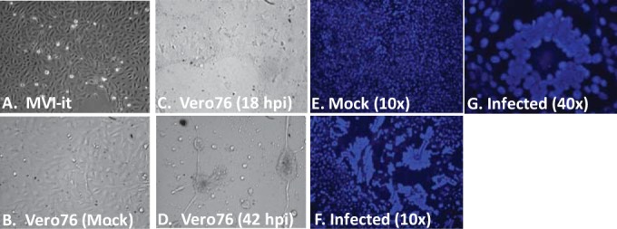
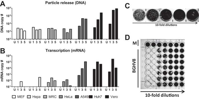
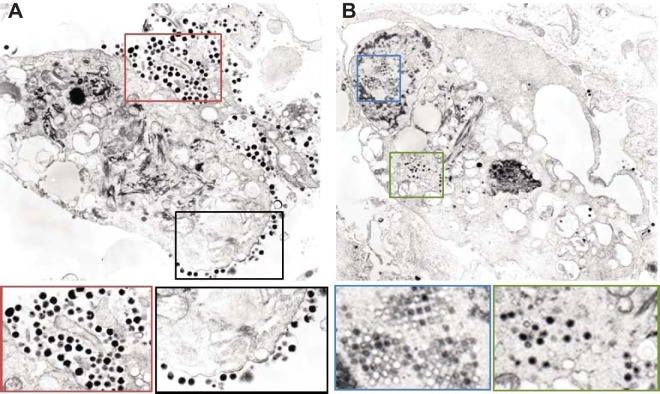
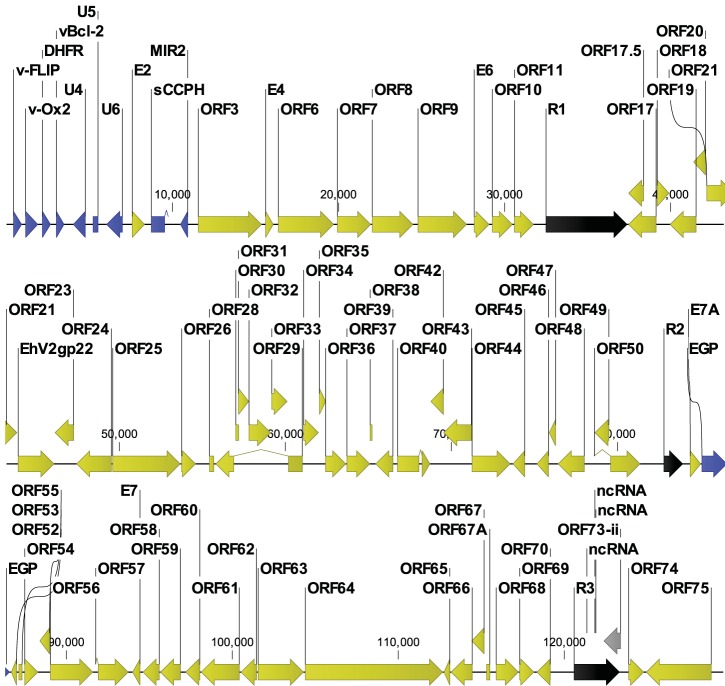
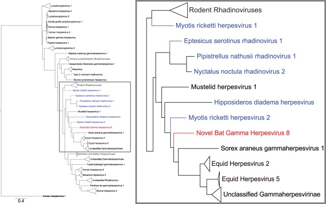
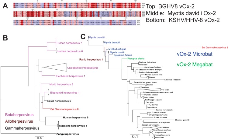
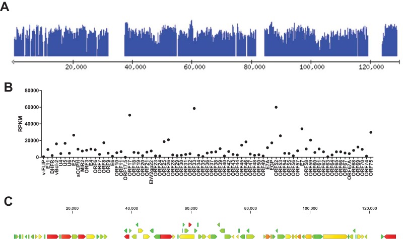
While employing deep sequencing and de novo assembly to characterize the mRNA transcript profile of a cell line derived from the microbat Myotis velifer incautus, we serendipitously identified mRNAs encoding proteins with a high level of identity to herpesviruses. A majority were closely related to proteins of equine herpesvirus 2 (EHV-2), a horse gammaherpesvirus. We demonstrated by electron microscopy the presence of herpesvirus-like particles in the microbat cells. Passage of supernatants from microbat cells to Vero cells resulted in syncytium formation, and expression of viral genes and amplification of viral DNA were demonstrated by quantitative PCR. Susceptibility of human cell lines to productive infection was also demonstrated. Next-generation sequencing and de novo assembly of the viral genome from supernatants from Vero cells yielded a single contig of approximately 130 kb with at least 77 open reading frames (ORFs), predicted microRNAs (miRNAs), and a gammaherpesvirus genomic organization. Phylogenic analysis of the envelope glycoprotein (gB) and DNA polymerase (POLD1) revealed similarity to multiple gammaherpesviruses, including those from as-yet-uncultured viruses of the Rhadinovirus genus that were obtained by deep sequencing of bat tissues. Moreover, the assembled genome revealed ORFs that share little or no homology to known ORFs in EHV-2 but are similar to accessory proteins of other gammaherpesviruses. Some also have striking homology to predicted Myotis bat proteins. Cumulatively, this study provides the first isolation and characterization of a replication-competent bat gammaherpesvirus. IMPORTANCE Bats are of significant interest as reservoirs for zoonotic viral pathogens; however, tools to dissect bat-virus interactions are limited in availability. This study serendipitously identified, in an established bat cell line, a fully replication-competent gammaherpesvirus; determined the complete genome sequence of the virus; and generated a viral transcript map. This virus can replicate in select human and nonhuman primate cell lines. However, analyses of viral sequences support a bat origin for this virus; we therefore refer to the virus as bat gammaherpesvirus 8 (BGHV8). The viral genome contains unique open reading frames that likely encode modulators of bat innate and adaptive immune signaling pathways and expresses viral miRNAs. The virus and its gene products should provide a unique tool to dissect both bat and gammaherpesvirus biology.

Keywords: bats; genomics; herpesviruses; next-generation sequencing; transcriptomics; virus discovery.


Figures

FIG 1
FIG 1
Supernatant transfer from the MVI-it bat cell line to Vero cells results in syncytia and CPE. (A) MVI-it cells growing in culture harboring the novel gammaherpesvirus. (B) Phase-contrast image of Vero cells prior to MVI-it supernatant transfer. (C) Phase-contrast image of Vero cells 18 h after MVI-it supernatant transfer. (D) Phase-contrast image of Vero cells 42 h after supernatant transfer. (E) Hoechst-stained Vero cells from panel B display nuclei prior to MVI-it supernatant transfer. (F and G) Hoechst-stained Vero cells 18 h after supernatant transfer display evidence of syncytia.
FIG 2
FIG 2
Quantitation of bat gammaherpesvirus 8-infected cells and release in supernatant from multiple cell lines. (A) Quantitative PCR (qPCR) to measure the amount of viral DNA released into the supernatant of seven representative cell lines at days 1, 3, and 5 postinfection. (B) Similar to panel A, but mRNA from infected cells was analyzed by reverse transcription followed by qPCR. (C) Representative plaque assay of BGHV8 on Vero cells. (D) Representative 50% tissue culture infectious dose (TCID50) data from BGHV8 on Vero cells.
FIG 3
FIG 3
Electron microscopy of the MVI-it cell line identifies viral particles that appear to be BGHV8. (A) Representative EM image from the MVI-it culture. The black and red boxes highlight enlarged regions directly below the image above. Both areas identify budding and fully budded herpesvirus virions. (B) Representative EM image from the MVI-it culture. The blue and green boxes highlight enlarged regions directly below the image above. Both areas highlight intracellular herpesvirus particles, and the blue box captures virions within an inclusion body. Courtesy of Vsevolod Popov and Krystle Agans, reproduced with permission.
FIG 4
FIG 4
Assembly of the novel bat gammaherpesvirus genome highlights ORFs related to EHV-2 and accessory ORFs related to other gammaherpesviruses. To assemble a single contig of 129,563 bp, contigs assembled with 100-bp reads from Illumina HiSeq were joined with 270-bp reads from Illumina MiSeq data. The genome was annotated using the Viral Genome ORF Reader (VIGOR). Open reading frames are highlighted as follows: yellow, ORFs with homology to EHV-2; blue, ORFs with homology to other herpesvirus proteins; black, repeat regions; gray, putative LANA gene with an internal unresolved repeat region. ncRNA, noncoding RNA.
FIG 5
FIG 5
gB protein phylogenic tree highlights similarities with other bat herpesviruses and EHV-2. The tree displays multiple gammaherpesvirus protein sequences, and human herpesvirus 1 represents the outgroup. BGHV8 gB is an 839-amino-acid protein. An alignment was generated for a 263-amino-acid region of gB from positions 498 to 760 of BGHV8. All sequences were trimmed to include only the corresponding region, and an alignment was derived with an LG substitution model and 100 bootstraps with a scale of 0.4. The inset on the right side displays an enlarged image highlighted corresponding to the gray box in the left image to display the sequences most closely related to BGHV8. Red denotes BGHV8, and blue labels denote gB from viruses obtained from bat species. The most closely related sequence from this analysis comes from a gB sequence of Myotis ricketti herpesvirus 2 (AFM85235.1). Triangles represent multiple sequences which were collapsed to enhance presentation. To see the tree with no collapsed sequences and corresponding accession numbers, see Fig. S1 in the supplemental material.
FIG 6
FIG 6
BGHV8 v-OX2 clusters with gammaherpesviruses and Myotis sequences. (A) Representative alignment of BGHV8 OX2 with KSHV v-OX2 and Myotis davidii OX2. Dark red indicates identical residues, pink indicates conservation between two of the three alignments, and blue indicates less conserved regions. (B) Tree of representative viral OX2 ORFs demonstrates the relationship of BGHV8 v-OX2 to other gammaherpesviruses. Bold black denotes the penguinpox virus v-OX2 outgroup, black indicates gammaherpesvirus v-OX2, and pink denotes betaherpesvirus v-OX2. The red label highlights the location of BGHV8. (C) Tree of representative mammalian OX2 ORFs demonstrates the relatedness of BGHV8 OX2 to those from bat species. Blue labels denote microbat sequences, and green labels denote megabat sequences. The red label highlights the location of BGHV8.
FIG 7
FIG 7
RNA-seq analysis from the MVI-it culture indicates that a majority of the BGHV8 coding sequences are transcribed. (A) Screen shot of RNA-seq reads mapped to each CDS of the BGHV8 genome. The data are presented in the Lightweight Genome Viewer (lwgv). The y axis is a log10 coverage scale, and the genomic position is displayed on the x axis. Interactive access to the coverage data across the BGHV8 genome is available at http://katahdin.mssm.edu/ravi/web/lwgv/lwgv.cgi?ann=nbghv_3.ann. (B) Reads per kilobase of transcript per million mapped reads (RPKM) for each CDS of BGHV8 are displayed. (C) A heat map imposed on the BGHV8 genome also illustrates the relative expression of each CDS. Green denotes the lowest RPKM values, yellow/orange indicates intermediate RPKM values, and red indicates the highest RPKM values. Raw reads and RPKM data are available through GEO (GSE76756).

References

    1. Jones KE, Patel NG, Levy MA, Storeygard A, Balk D, Gittleman JL, Daszak P. 2008. Global trends in emerging infectious diseases. Nature 451:990–993. doi: 10.1038/nature06536. - DOI - PMC - PubMed
    1. Teeling EC, Springer MS, Madsen O, Bates P, O’Brien SJ, Murphy WJ. 2005. A molecular phylogeny for bats illuminates biogeography and the fossil record. Science 307:580–584. doi: 10.1126/science.1105113. - DOI - PubMed
    1. Wang L-F, Cowled C. 2015. Bats and viruses: a new frontier of emerging infectious diseases. Wiley-Blackwell, Hoboken, NJ.
    1. Sunagawa S, Coelho LP, Chaffron S, Kultima JR, Labadie K, Salazar G, Djahanschiri B, Zeller G, Mende DR, Alberti A, Cornejo-Castillo FM, Costea PI, Cruaud C, d’Ovidio F, Engelen S, Ferrera I, Gasol JM, Guidi L, Hildebrand F, Kokoszka F, Lepoivre C, Lima-Mendez G, Poulain J, Poulos BT, Royo-Llonch M, Sarmento H, Vieira-Silva S, Dimier C, Picheral M, Searson S, Kandels-Lewis S, Bowler C, de Vargas C, Gorsky G, Grimsley N, Hingamp P, Iudicone D, Jaillon O, Not F, Ogata H, Pesant S, Speich S, Stemmann L, Sullivan MB, Weissenbach J, Wincker P, Karsenti E, Raes J, Acinas SG, Bork P. 2015. Ocean plankton. Structure and function of the global ocean microbiome. Science 348:1261359. doi: 10.1126/science.1261359. - DOI - PubMed
    1. Bikel S, Valdez-Lara A, Cornejo-Granados F, Rico K, Canizales-Quinteros S, Soberon X, Del Pozo-Yauner L, Ochoa-Leyva A. 2015. Combining metagenomics, metatranscriptomics and viromics to explore novel microbial interactions: towards a systems-level understanding of human microbiome. Comput Struct Biotechnol J 13:390–401. doi: 10.1016/j.csbj.2015.06.001. - DOI - PMC - PubMed