. 2016 Mar 30;1(2):e00007-16.
doi: 10.1128/mSphere.00007-16. eCollection 2016 Mar-Apr.

A Single Residue in Ebola Virus Receptor NPC1 Influences Cellular Host Range in Reptiles

Affiliations

A Single Residue in Ebola Virus Receptor NPC1 Influences Cellular Host Range in Reptiles

Esther Ndungo et al. mSphere. .

Abstract

Filoviruses are the causative agents of an increasing number of disease outbreaks in human populations, including the current unprecedented Ebola virus disease (EVD) outbreak in western Africa. One obstacle to controlling these epidemics is our poor understanding of the host range of filoviruses and their natural reservoirs. Here, we investigated the role of the intracellular filovirus receptor, Niemann-Pick C1 (NPC1) as a molecular determinant of Ebola virus (EBOV) host range at the cellular level. Whereas human cells can be infected by EBOV, a cell line derived from a Russell's viper (Daboia russellii) (VH-2) is resistant to infection in an NPC1-dependent manner. We found that VH-2 cells are resistant to EBOV infection because the Russell's viper NPC1 ortholog bound poorly to the EBOV spike glycoprotein (GP). Analysis of panels of viper-human NPC1 chimeras and point mutants allowed us to identify a single amino acid residue in NPC1, at position 503, that bidirectionally influenced both its binding to EBOV GP and its viral receptor activity in cells. Significantly, this single residue change perturbed neither NPC1's endosomal localization nor its housekeeping role in cellular cholesterol trafficking. Together with other recent work, these findings identify sequences in NPC1 that are important for viral receptor activity by virtue of their direct interaction with EBOV GP and suggest that they may influence filovirus host range in nature. Broader surveys of NPC1 orthologs from vertebrates may delineate additional sequence polymorphisms in this gene that control susceptibility to filovirus infection. IMPORTANCE Identifying cellular factors that determine susceptibility to infection can help us understand how Ebola virus is transmitted. We asked if the EBOV receptor Niemann-Pick C1 (NPC1) could explain why reptiles are resistant to EBOV infection. We demonstrate that cells derived from the Russell's viper are not susceptible to infection because EBOV cannot bind to viper NPC1. This resistance to infection can be mapped to a single amino acid residue in viper NPC1 that renders it unable to bind to EBOV GP. The newly solved structure of EBOV GP bound to NPC1 confirms our findings, revealing that this residue dips into the GP receptor-binding pocket and is therefore critical to the binding interface. Consequently, this otherwise well-conserved residue in vertebrate species influences the ability of reptilian NPC1 proteins to bind to EBOV GP, thereby affecting viral host range in reptilian cells.

Keywords: Ebola virus; NPC1; Niemann-Pick C1; endosomal receptor; filovirus; intracellular receptor; reptiles; viral receptor; virus-host interactions.


Figures

FIG 1
FIG 1
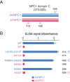
Alignment of human and viper NPC1 domains C. (A) Schematic of full-length NPC1 protein, showing luminal domains A, C, and I. (B) Alignment of NPC1 domain C sequences from human and Russell’s viper. Cysteine residues are in blue. Predicted N-glycosylation sites (sequons) that are conserved in the two proteins are indicated with black arrowheads. Orange arrowheads mark those unique to Russell’s viper NPC1 domain C, and a green arrowhead marks one that is unique to human NPC1 domain C. Nonidentical residues are highlighted in yellow. Position 503 is highlighted in pink.
FIG 2
FIG 2
Both HsNPC1 and DrNPC1 domain C proteins are expressed and secreted but bind differentially to EBOV GPCL. (A) Soluble forms of the NPC1 domain C proteins from human (HsNPC1) and Russell’s viper (DrNPC1) were expressed in FreeStyle 293-F cells and purified by nickel-affinity chromatography. Equal concentrations were resolved by anti-Flag immunostaining. Left, no treatment. Right, treatment with protein N-glycosidase F (PNGase F). Numbers at left are molecular masses in kilodaltons, and numbers at right are relative molecular weights in thousands. (B) The two NPC1 domain C proteins were tested in an ELISA for binding to EBOV GPCL. VSV-EBOV GP viruses were cleaved with thermolysin (250 µg/ml) and captured on an ELISA plate using monoclonal antibody KZ52. Serial dilutions of either HsNPC1 or DrNPC1 domain C proteins were added, and binding to GPCL was detected by anti-Flag antibody.
FIG 3
FIG 3
HsNPC1-DrC chimera is functional at cholesterol clearance from lysosomes but does not support EBOV entry and infection. (A) Full-length NPC1 constructs—human WT (HsNPC1) and the human NPC1 chimera with the domain C replaced with viper domain C (HsNPC1-DrC)—immunostained with anti-Flag antibody (red) colocalize with the lysosomal marker LAMP1 (green) when transiently expressed in U2OS NPC1−/− cells (27). (B) CHO-M12 cells stably expressing either HsNPC1 WT or HsNPC1-DrC were stained with filipin to visualize unesterified cholesterol. Top panel, filipin staining. Cholesterol-laden cells are marked with red arrowheads. Blue arrowheads indicate cells that are functional at cholesterol clearance. Bottom panel, cells immunostained with anti-Flag antibody for NPC1 expression (green). (C) Infection of cells from panel B by authentic EBOV (multiplicity of infection of 10), scored 72 h postinfection and normalized to infection on HsNPC1(WT). (D) Infection of cells from panel B by VSV-EBOV GP calculated by manual counting of eGFP-positive cells. IU/ml, infectious units per milliliter. Means ± standard deviations (n = 2 to 4) from a representative experiment are shown in each panel.
FIG 4
FIG 4
N-glycosylation of NPC1 domain C does not affect EBOV GPCL binding. (A) Location of the three unique sequons in HsNPC1 versus DrNPC1 domain C. (B) Glycosylation mutants were made in both HsNPC1 (losing sequon at position 598 and gaining sequons at position 414 and 498) and DrNPC1 (losing sequons at position 414 and 498 and gaining sequon at position 598). Domain C proteins were expressed in HEK 293T cells and tested for EBOV GPCL binding by ELISA.
FIG 5
FIG 5
Middle region of HsNPC1 domain C confers binding ability on DrNPC1. (A) Chimeras were engineered by replacing DrNPC1 domain C sequences with human sequence 373 to 475 (chimera 1), 476 to 536 (chimera 2), or 537 to 620 (chimera 3). The chimeras were expressed in HEK 293T cells and tested for EBOV GPCL binding by ELISA. (B) Further dissection of chimera 2 was done by replacing smaller subsets of DrNPC1 with human residues 494 to 502 (chimera 4), 502 to 511 (chimera 5), and 527 to 531 (chimera 6). Chimeric NPC1 domain C proteins were tested as in panel A.
FIG 6
FIG 6
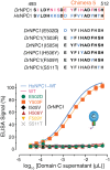
A single amino acid change renders DrNPC1 domain C fully competent to bind EBOV GPCL. Chimera 5 contains 6 amino acid differences between DrNPC1 and HsNPC1 domain C. The following point mutations were made in the DrNPC1 domain C by switching the viper amino acid residue at each of these positions to the corresponding human residue: E502D, Y503F, I505V, H506Y, F509Y, and S511T. The point mutants were expressed in HEK 293T cells and tested for EBOV GPCL binding by ELISA.
FIG 7
FIG 7
NPC1 residue 503 bidirectionally alters domain C’s capacity to bind EBOV GPCL. (A) HsNPC1 and DrNPC1 domain C proteins bearing point mutations at residue 503 (HsNPC1, F503Y; DrNPC1, Y503F) were expressed and purified. (B) Serial dilutions of equivalent amounts of purified NPC1 domain C proteins were tested for EBOV GPCL binding by ELISA.
FIG 8
FIG 8
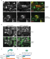
Residue 503 influences the capacity of full-length NPC1 to support EBOV entry and infection. (A) Point mutations at residue 503 were introduced into HsNPC1 and the chimera HsNPC1-DrC (F503Y and Y503F, respectively), and these constructs were transiently expressed in U2OS NPC1−/− cells. NPC1 (red) and a lysosomal marker, LAMP1 (green), were visualized by immunofluorescence microscopy. (B) NPC1-deficient CHO-M12 cells stably expressing either HsNPC1 (F503Y) or HsNPC1-DrC (Y503F) were stained with filipin to visualize unesterified cholesterol. Top panel, filipin staining. Bottom panel, cells immunostained with anti-Flag antibody for NPC1 expression (green). Cholesterol-laden cells are marked with red arrowheads. Blue arrowheads indicate cells that are functional at cholesterol clearance. (C) CHO-M12 cells stably expressing the NPC1 proteins indicated were exposed to authentic virus (multiplicity of infection of 3), scored at 72 h postinfection, and normalized to HsNPC1 WT infectivity. (D) Infection by VSV-EBOV GP, calculated by manual counting of eGFP-positive cells. IU/ml, infectious units per milliliter. Means ± standard deviations (n = 4) from a representative experiment are shown in each panel.
FIG 9
FIG 9
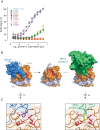
A bulky, hydrophobic residue is required at position 503. (A) The F at NPC1 residue 503 mutated to A, D, H, L, S, T, Y, or W and tested for binding to EBOV GPCL by ELISA. (B) Structure of EBOV GP monomer with GP1 (orange) and GP2 (gray) and the glycan cap (blue) occluding the NPC1-binding site (residues identified as critical for binding are colored red) (PDB identifier 3CSY [28]). Proteolytic removal of the glycan cap and mucin domain (not shown) in host cell endosomes unmasks this site, allowing binding of NPC1 domain C (green) (PDB identifier 5F1B [26]). (C) Comparison of the interaction between residues W86, F88, L111, I113, L122, and V141 (red) in the GP1 hydrophobic trough and F225 (magenta) of the glycan cap (PDB identifier 3CSY [28]), left, versus F503 (yellow) of NPC1 domain C (PDB identifier 5F1B [26]), right.
FIG 10
FIG 10
The tyrosine residue at NPC1 position 503 is unique to the Russell’s viper and king cobra NPC1 orthologs. (A) Alignment of sequences flanking residue 503 (red arrowhead) in domain C from divergent NPC1 orthologs. Residues different from the human sequence are highlighted in yellow. F503 is shaded blue, and Y503 is shaded pink. (B and C) Binding of NPC1 domain C proteins from snakes (B) and other reptiles (C) to EBOV GPCL as determined by ELISA.

References

    1. Baize S, Pannetier D, Oestereich L, Rieger T, Koivogui L, Magassouba N, Soropogui B, Sow MS, Keïta S, De Clerck H, Tiffany A, Dominguez G, Loua M, Traoré A, Kolié M, Malano ER, Heleze E, Bocquin A, Mély S, Raoul H, Caro V, Cadar D, Gabriel M, Pahlmann M, Tappe D, Schmidt-Chanasit J, Impouma B, Diallo AK, Formenty P, Van Herp M, Günther S. 2014. Emergence of Zaire Ebola virus disease in Guinea. N Engl J Med 371:1418–1425. doi:10.1056/NEJMoa1404505. - DOI - PubMed
    1. de La Vega M-A, Stein D, Kobinger GP. 2015. Ebolavirus evolution: past and present. PLoS Pathog 11:e1005221. doi:10.1371/journal.ppat.1005221. - DOI - PMC - PubMed
    1. Negredo A, Palacios G, Vázquez-Morón S, González F, Dopazo H, Molero F, Juste J, Quetglas J, Savji N, de la Cruz Martínez M, Herrera JE, Pizarro M, Hutchison SK, Echevarría JE, Lipkin WI, Tenorio A. 2011. Discovery of an Ebolavirus-like filovirus in Europe. PLoS Pathog 7:e1002304. doi:10.1371/journal.ppat.1002304. - DOI - PMC - PubMed
    1. Leroy EM, Kumulungui B, Pourrut X, Rouquet P, Hassanin A, Yaba P, Délicat A, Paweska JT, Gonzalez J-P, Swanepoel R. 2005. Fruit bats as reservoirs of Ebola virus. Nature 438:575–576. doi:10.1038/438575a. - DOI - PubMed
    1. Towner JS, Pourrut X, Albariño CG, Nkogue CN, Bird BH, Grard G, Ksiazek TG, Gonzalez J-P, Nichol ST, Leroy EM. 2007. Marburg virus infection detected in a common African bat. PLoS One 2:e764. doi:10.1371/journal.pone.0000764. - DOI - PMC - PubMed