. 2016 Jun;72(Pt 6):434-42.
doi: 10.1107/S2053230X16007202. Epub 2016 May 23.

Cyclization strategies of meditopes: affinity and diffraction studies of meditope-Fab complexes

Affiliations

Cyclization strategies of meditopes: affinity and diffraction studies of meditope-Fab complexes

Krzysztof P Bzymek et al. Acta Crystallogr F Struct Biol Commun. 2016 Jun.

Abstract

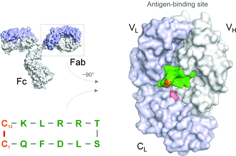
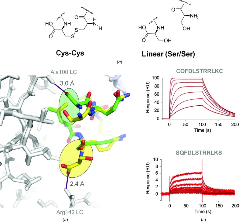
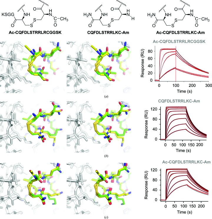
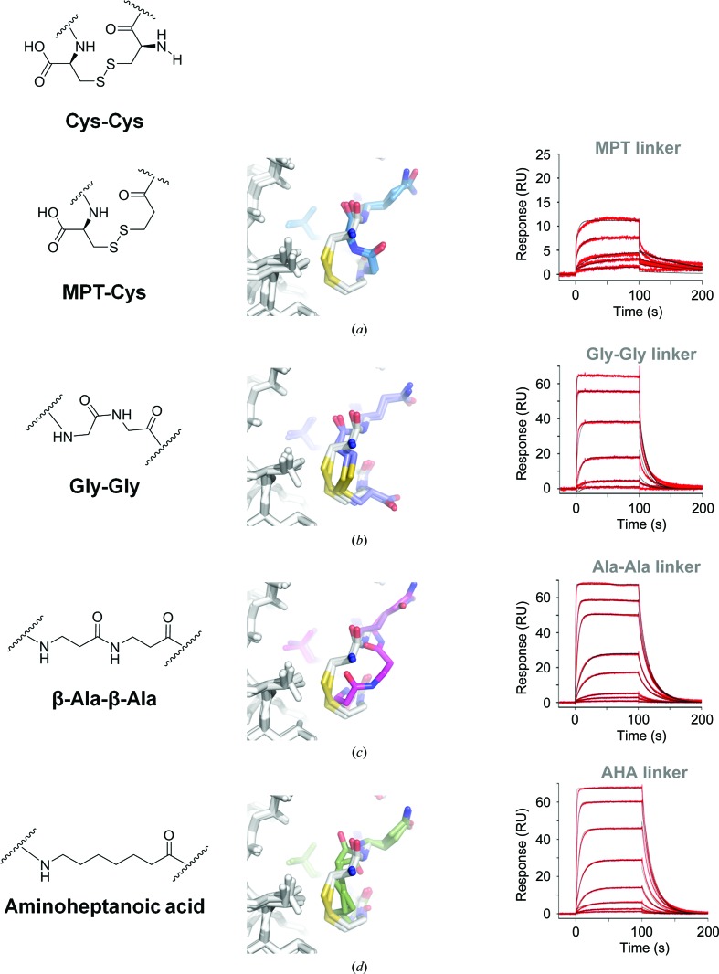
Recently, a unique binding site for a cyclic 12-residue peptide was discovered within a cavity formed by the light and heavy chains of the cetuximab Fab domain. In order to better understand the interactions that drive this unique complex, a number of variants including the residues within the meditope peptide and the antibody, as well as the cyclization region of the meditope peptide, were created. Here, multiple crystal structures of meditope peptides incorporating different cyclization strategies bound to the central cavity of the cetuximab Fab domain are presented. The affinity of each cyclic derivative for the Fab was determined by surface plasmon resonance and correlated to structural differences. Overall, it was observed that the disulfide bond used to cyclize the peptide favorably packs against a hydrophobic `pocket' and that amidation and acetylation of the original disulfide meditope increased the overall affinity ∼2.3-fold. Conversely, replacing the terminal cysteines with serines and thus creating a linear peptide reduced the affinity over 50-fold, with much of this difference being reflected in a decrease in the on-rate. Other cyclization methods, including the formation of a lactam, reduced the affinity but not to the extent of the linear peptide. Collectively, the structural and kinetic data presented here indicate that small perturbations introduced by different cyclization strategies can significantly affect the affinity of the meditope-Fab complex.

Keywords: X-ray crystallography; meditope; monoclonal antibody; surface plasmon resonance.


Figures

Figure 1
Figure 1
Fab–meditope interface. The light chain is shown in light blue, the heavy chain in dark blue and the meditope in green; the linker residues subjected to mutagenesis are highlighted in orange (highlighted by a red arrow).
Figure 2
Figure 2
Cyclic versus acyclic. (a) Replacement of Cys1 and Cys12 with serine as a conservative substitution to create a linear meditope. (b) There are two Fab–peptide complexes in the asymmetric unit. Superposition of these complexes indicates flexibility at the N-termini. In one peptide Fab–complex (the peptide with yellow C atoms), the terminal carboxylate makes a favorable salt bridge with the Arg142 guanidinium group from the light chain (LC). In the other peptide–Fab complex (the peptide with green C atoms), the hydroxyl group of Ser1 makes a hydrogen bond to the carbonyl group of Ala100, also of the light chain. However, the electron density is weak and Ser12 could not be built. (c) SPR traces of the original meditope and the linear meditope indicate a substantial reduction in binding affinity and an increase in the off-rate.
Figure 3
Figure 3
Terminal modifications. Each Fab–peptide complex was superimposed and is shown in stereo with the corresponding SPR trace. (a) Long meditope, (b) amidated, (c) acetylated and amidated complexes. In each case the disulfide packs against Val9 and Ile10 of the light chain. Eliminating charge in each case did not produce substantial structural differences, but did improve the overall affinity.
Figure 4
Figure 4
Alternative linking strategies. The left column indicates the modification. The middle column shows the superposition of each Fab–alternative cyclic peptide complex (colored C atoms) on the Fab–acetylated-amidated disulfide peptide (white C atoms). The right column is the corresponding SPR trace. In all cases, the linking atoms are shifted away from the Val9/Ile10 residues and thus do not pack as well. The affinity of the aminoheptanoic acid linker (AHA) was the closest to the original disulfide meditope.
Figure 5
Figure 5
Superposition of all meditope peptide variants. Superposition of each peptide variant (colored by thermal factor) from each complex indicates that the ‘core’ residues, amino acids 3–10, are effectively not perturbed by the cyclization strategies.

References

    1. Adams, P. D. et al. (2010). Acta Cryst. D66, 213–221. - PubMed
    1. Donaldson, J. M., Zer, C., Avery, K. N., Bzymek, K. P., Horne, D. A. & Williams, J. C. (2013). Proc. Natl Acad. Sci. USA, 110, 17456–17461. - PMC - PubMed
    1. Emsley, P., Lohkamp, B., Scott, W. G. & Cowtan, K. (2010). Acta Cryst. D66, 486–501. - PMC - PubMed
    1. Herbst, R. S., Kim, E. S. & Harari, P. M. (2001). Expert Opin. Biol. Ther. 1, 719–732. - PubMed
    1. Huang, S.-M., Bock, J. M. & Harari, P. M. (1999). Cancer Res. 59, 1935–1940. - PubMed

Publication types

Substances