. 2016 Jun;22(2):296-307.
doi: 10.3350/cmh.2016.0028.

Liver imaging reporting and data system (LI-RADS) version 2014: understanding and application of the diagnostic algorithm

Affiliations

Liver imaging reporting and data system (LI-RADS) version 2014: understanding and application of the diagnostic algorithm

Chansik An et al. Clin Mol Hepatol. 2016 Jun.

Abstract

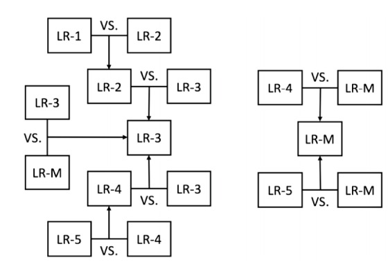
Liver Imaging Reporting and Data System (LI-RADS) is a system for interpreting and reporting of computed tomography and magnetic resonance imaging of the liver in patients at risk for hepatocellular carcinoma (HCC). LI-RADS has been developed to address the limitations of prior imaging-based criteria including the lack of established consensus regarding the exact definitions of imaging features, binary categorization (either definite or not definite HCC), and failure to consider non-HCC malignancies. One of the most important goals of LI-RADS is to facilitate clear communication between all the personnel involved in the diagnosis and treatment of HCC, such as radiologists, hepatologists, surgeons, and pathologists. Therefore, clinicians should also be familiar with LI-RADS. This article reviews the LI-RADS diagnostic algorithm, and the definitions and management implications of LI-RADS categories.

Keywords: Algorithms; Carcinoma; Diagnosis; Guideline; Hepatocellular.


Conflict of interest statement

Conflicts of Interest: The authors have no conflicts to disclose.

Figures

Figure 1.
Figure 1.
Summary schematic of LI-RADS 2014 diagnostic algorithm (adapted and modified from ACR LI-RADS content at: http://www.acr.org/Quality-Safety/Resources/LIRADS). LI-RADS, liver imaging reporting and data system; ACR, American College of Radiology; LR, LI-RADS. *Observations in this cell are categorized as LR-4 except as follows: LR-5g, if there is ≥50% diameter increase in ≤6 months, and LR-5us, if there is both “washout” and visibility as discrete nodules at antecedent surveillance ultrasound.
Figure 2.
Figure 2.
An observation that can be categorized as LR-2. At gadoxetic acid-enhanced MRI, a hepatic observation in the segment 5 of liver is distinguished from background liver as a 1.7 cm hyperintense nodule on pre-contrast T1-weighted image (arrow) (A), but shows no hyperenhancement in the arterial phase (B), no washout appearance in the portal venous phase (C), and iso-intensity on T2-weighted image (D).
Figure 3.
Figure 3.
An observation that can be categorized as LR-3. At gadoxetic acid-enhanced MRI, a 1.2 cm nodule in the hepatic dome shows no arterial hyperenhancement (A). However, this observation would be better categorized as LR-3 than as LR-2, because of the presence of washout appearance in the portal venous phase (B) and some ancillary features such as hepatobiliary phase hypointensity (C) and moderate T2 hyperintensity (D).
Figure 4.
Figure 4.
An observation that can be categorized as LR-4, LR-5us, or LR-5g depending on the clinical situation. In the arterial phase of gadoxetic acid-enhanced MRI (arrow) (A), this 1.2 cm subcapsular lesion in the segment 7 of liver shows hyperenhancement, which is more definite in the subtraction image (arrow) (B), which is more definite in the subtraction image (B). In the portal venous (C) and delayed (D) phases, the lesion shows washout appearance but no capsule appearance. Because the nodule is smaller than 2 cm, this lesion cannot be categorized as LR-5 despite the presence of the radiologic hallmark of hepatocellular carcinoma (arterial hyperenhancement and washout). However, this 1- to 2-cm nodule with the radiologic hallmark can be categorized as LR-5, if it was previously detected at US (LR-5us) or showed ≥50% diameter increase in ≤6 months (LR-5g).
Figure 5.
Figure 5.
An observation that can be categorized as LR-5. In the pre-contrast T1-weighted image (A) and arterial (B), portal venous (C), and delayed (D) phases of gadoxetic acid-enhanced MRI, this 1.3 cm nodule unequivocally shows arterial hyperenhancement (B), washout appearance (C), and capsule appearance (C and D).
Figure 6.
Figure 6.
An observation that can be categorized as LR-5V. In the pre-contrast (A), arterial (B), portal venous (C), and delayed (D) phases of dynamic liver CT, ill-defined infiltrative lesions are diffusely involving the right posterior hepatic section (arrow), with expansile tumor thrombi in both main portal vein branches (arrowheads).
Figure 7.
Figure 7.
An observation that can be categorized as LR-M. At gadopentetate-enhanced MRI, a 4-cm mass in the segment 5 of liver shows irregular peripheral enhancement and peritumoral parenchymal enhancement in the arterial phase (A) and peripheral washout and central enhancement in the delayed phase (B). These imaging features favor other malignancy over hepatocellular carcinoma (HCC).
Figure 8.
Figure 8.
Tie breaking rules to adjust category. If still unsure about the final category for an observation after application of ancillary features, these tie-breaking rules should be applied. LR, LI-RADS.

References

    1. Omata M, Lesmana LA, Tateishi R, Chen PJ, Lin SM, Yoshida H, et al. Asian Pacific Association for the Study of the Liver consensus recommendations on hepatocellular carcinoma. Hepatol Int. 2010;4:439–474. - PMC - PubMed
    1. Bruix J, Sherman M. Management of hepatocellular carcinoma: an update. Hepatology. 2011;53:1020–1022. - PMC - PubMed
    1. EASL-EORTC clinical practice guidelines: management of hepatocellular carcinoma. J Hepatol. 2012;56:908–943. - PubMed
    1. Wald C, Russo MW, Heimbach JK, Hussain HK, Pomfret EA, Bruix J. New OPTN/UNOS policy for liver transplant allocation: standardization of liver imaging, diagnosis, classification, and reporting of hepatocellular carcinoma. Radiology. 2013;266:376–382. - PubMed
    1. Kudo M, Matsui O, Izumi N, Iijima H, Kadoya M, Imai Y. Surveillance and diagnostic algorithm for hepatocellular carcinoma proposed by the Liver Cancer Study Group of Japan: 2014 update. Oncology. 2014;87:S7–S21. - PubMed