. 2016 Jun 15;12(6):e1006102.
doi: 10.1371/journal.pgen.1006102. eCollection 2016 Jun.

Dbl2 Regulates Rad51 and DNA Joint Molecule Metabolism to Ensure Proper Meiotic Chromosome Segregation

Affiliations

Dbl2 Regulates Rad51 and DNA Joint Molecule Metabolism to Ensure Proper Meiotic Chromosome Segregation

Silvia Polakova et al. PLoS Genet. .

Abstract

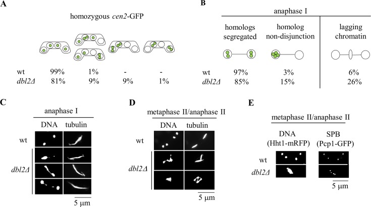
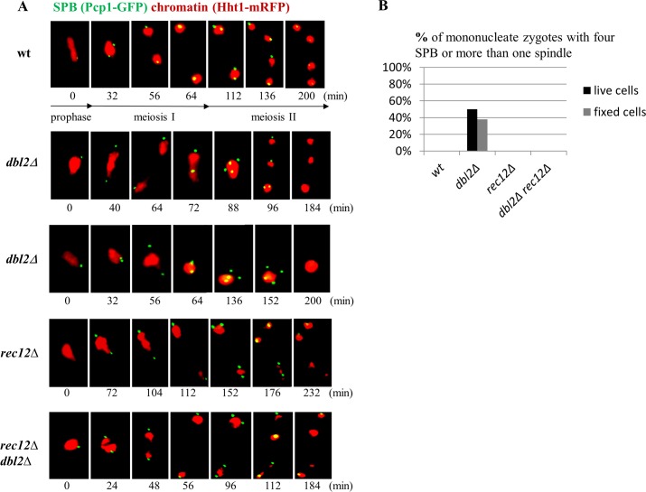
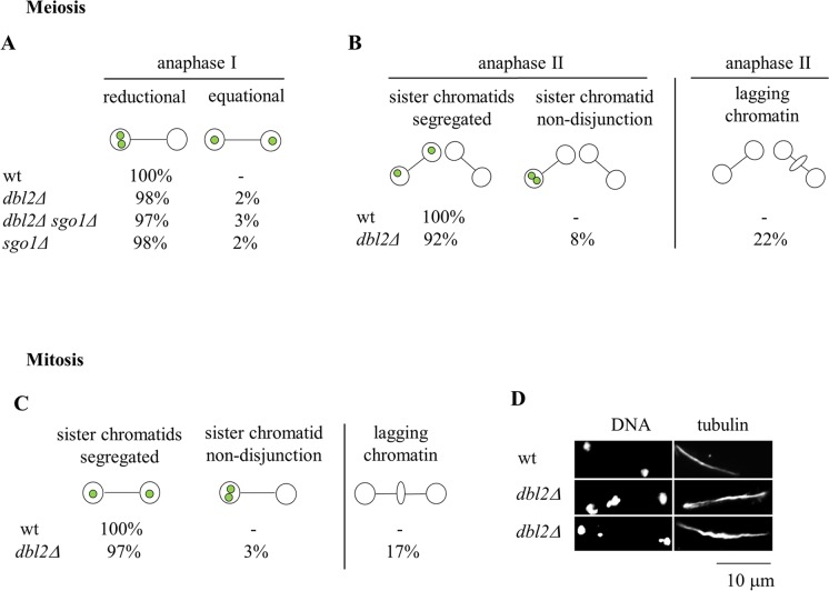
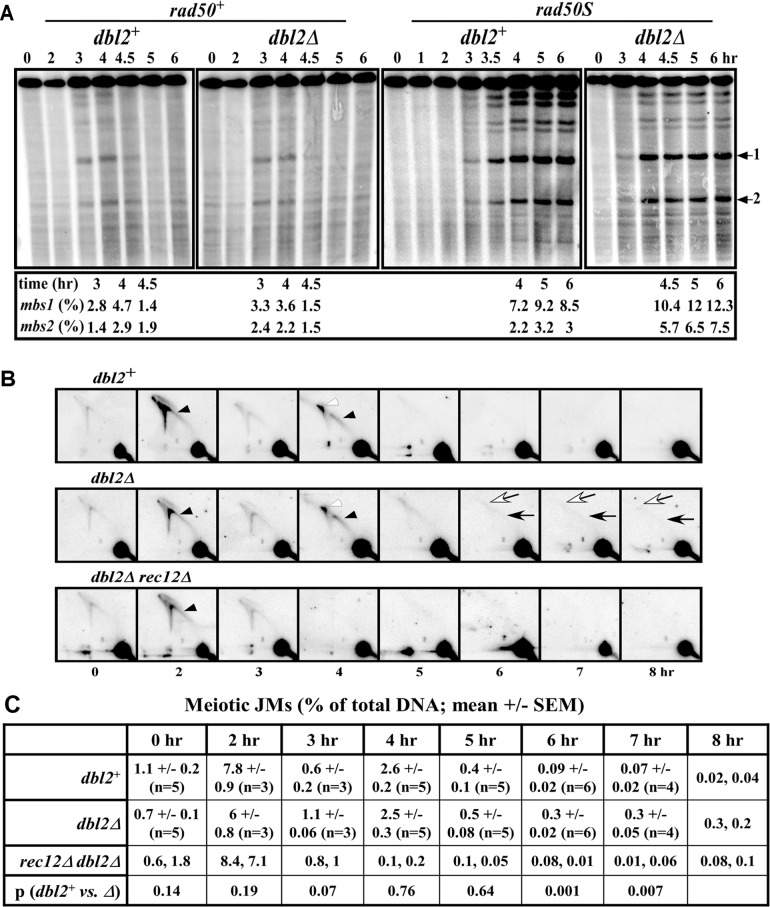
To identify new proteins required for faithful meiotic chromosome segregation, we screened a Schizosaccharomyces pombe deletion mutant library and found that deletion of the dbl2 gene led to missegregation of chromosomes during meiosis. Analyses of both live and fixed cells showed that dbl2Δ mutant cells frequently failed to segregate homologous chromosomes to opposite poles during meiosis I. Removing Rec12 (Spo11 homolog) to eliminate meiotic DNA double-strand breaks (DSBs) suppressed the segregation defect in dbl2Δ cells, indicating that Dbl2 acts after the initiation of meiotic recombination. Analyses of DSBs and Holliday junctions revealed no significant defect in their formation or processing in dbl2Δ mutant cells, although some Rec12-dependent DNA joint molecules persisted late in meiosis. Failure to segregate chromosomes in the absence of Dbl2 correlated with persistent Rad51 foci, and deletion of rad51 or genes encoding Rad51 mediators also suppressed the segregation defect of dbl2Δ. Formation of foci of Fbh1, an F-box helicase that efficiently dismantles Rad51-DNA filaments, was impaired in dbl2Δ cells. Our results suggest that Dbl2 is a novel regulator of Fbh1 and thereby Rad51-dependent DSB repair required for proper meiotic chromosome segregation and viable sex cell formation. The wide conservation of these proteins suggests that our results apply to many species.


Conflict of interest statement

The authors have declared that no competing interests exist.

Figures

Fig 1
Fig 1. Dbl2 is required for proper segregation of chromosomes during meiosis I.
(A) The segregation of chromosome 2 was scored in wild-type (JG12618) and dbl2Δ (JG17130) h90 strains in which chromosome 2 was marked with GFP (cen2-GFP). The strains were induced for meiosis and at 20–24 hr fixed and DNA was visualized by Hoechst staining. Chromosome 2 segregation (cen2-GFP dots) was scored in 500 asci. (B) The strains described in (A) were immunostained for tubulin and GFP; DNA was visualized by DAPI. cen2-GFP dots and lagging chromatin were scored under the fluorescence microscope in 100 anaphase I cells. (C) Examples of anaphase I zygotes in wild-type and dbl2Δ mutant, showing lagging chromatin and unsegregated DNA. (D) The images show spindle and chromatin morphology at metaphase II or anaphase II in wild type (JG15456 x JG11318) and dbl2Δ mutant (JG17208 x JG17207) meiosis. Cells were fixed and immunostained for tubulin; DNA was visualized by Hoechst staining. Mononucleate dbl2Δ zygotes with two or three spindles are shown. (E) Spindle pole bodies (SPBs) and chromosomes were visualized by endogenously tagged Pcp1-GFP and Hht1-mRFP, respectively. In a wild-type strain (JG16917), four SPBs are present at the completion of meiosis II, and each is associated with one of the four nuclei. In the dbl2Δ mutant (JG17116), mononucleate zygotes containing up to four SPBs are observed.
Fig 2
Fig 2. Deletion of dbl2 causes a Rec12-dependent failure of chromosome segregation during meiosis I.
(A) A wild-type strain (JG16917), dbl2Δ mutant (JG17116), rec12Δ mutant (JG17159) and rec12Δ dbl2Δ double mutant (JG17156) were plated on SPA sporulation plates and analyzed by live-cell imaging. The SPBs and chromosomes were observed via endogenously tagged Pcp1-GFP and Hht1-mRFP, respectively. Numbers below the images represent time, in minutes, elapsed since filming began at the end of the horsetail stage. Prophase, meiosis I and meiosis II are indicated. (B) The graph shows the percentage of live cells with four SPBs or more than one spindle on a single DNA mass (examples of such zygotes are shown in panel A) and the percentage of fixed cells with two or more spindles on a single DNA mass (examples of such zygotes are shown in Fig 1D). In live-cell imaging, 20 wild-type, 26 dbl2Δ, 10 rec12Δ and 17 dbl2Δ rec12Δ zygotes were scored. For the analysis of fixed cells, the strains (JG12618, JG17130, JG17351, JG17353) were immunostained for tubulin, DNA was visualized by Hoechst staining, and 100 zygotes from each strain were scored.
Fig 3
Fig 3. Dbl2 is required for proper segregation of sister chromatids during meiosis II and mitosis.
(A) h+ strains carrying cen2-GFP that were either wild-type (JG15456), dbl2Δ (JG17208), sgo1Δ (JG12269) or dbl2Δ sgo1Δ (JG17779) were crossed to h- strains of the same genotype but lacking cen2-GFP (JG11318, JG17207, JG11793 and JG17780). Cells were fixed and immunostained for tubulin and GFP; DNA was visualized by Hoechst staining. The segregation of chromosome 2 was scored in 100 anaphase I cells. (B) h+ cen2-GFP strains either wild-type (JG15456) or dbl2Δ (JG17208), were crossed to h- strains of the same genotype but lacking cen2-GFP (JG11318, JG17207). Zygotes were fixed and processed as in (A). The segregation of chromosome 2 and lagging chromatin were scored in 100 anaphase II cells. (C) The segregation of chromosome 2 was scored in mitotically dividing wild-type (JG15456) and dbl2Δ mutant (JG17208) cells. Cells were fixed and processed as in (A). The segregation of chromosome 2 and lagging chromatin were scored in 100 anaphase cells. (D) Examples of a wild-type anaphase cell and two dbl2Δ mutant cells showing lagging chromatin during anaphase.
Fig 4
Fig 4. DNA double-strand breaks and Holliday junctions are formed and repaired similarly in wild-type and dbl2Δ mutant, but Rec12-dependent joint molecules persist in dbl2Δ late meiosis.
(A) Formation and repair of DSBs on the 0.5 Mb NotI fragment J. Strains GP6656 (dbl2+), GP8664 (dbl2Δ) and GP8450 (dbl2Δ rad50S) were induced for meiosis; DNA was prepared at the indicated times, digested with NotI, and analyzed by pulsed-field gel electrophoresis and Southern blot hybridization using a probe at the left end of the 501 kb NotI fragment J (band at the top of the gel) [42]. GP3718 (dbl2+ rad50S) is from [94]. The fraction of total DNA broken at mbs1 and at mbs2 (indicated by arrows on the right) at the indicated time for each strain is shown below each blot. The timing of appearance and disappearance of DSBs and their frequencies in both rad50+ and rad50S strains are similar to those previously reported in wild-type cells [42,95]. (B) Formation and resolution of DNA joint molecules at the mbs1 hotspot. Strains GP6656 (dbl2+), GP8664 (dbl2Δ), and GP8836 (dbl2Δ rec12Δ) were induced for meiosis; DNA was extracted at the indicated times, digested with BsrGI, and analyzed by two-dimensional gel electrophoresis and Southern blot hybridization using a probe (~1 kb long) near the mbs1 DSB hotspot on the 10.5 kb BsrGI fragment (major spot at the bottom right) [96]. Black and white arrowheads, respectively, indicate Y-arc and X-spike species (replication and recombination intermediates) found transiently in wild-type cells. Black and white arrows indicate Y-arc and X-spike species, respectively, that persist only in dbl2Δ rec12+ cells (at 6, 7, and 8 hr). (C) Quantification of branched DNA (JMs) in panel B. ImageQuant analysis was used to quantify the amount of branched DNA (Y-arc plus X-spike) relative to the total DNA. Data are the mean ± SEM from n assays (dbl2+ and dbl2Δ) or individual data from duplicate assays (rec12Δ dbl2Δ). See S2 Table for values of all individual data. p values, from unpaired t-tests, are the probability that dbl2+ and dbl2Δ do not differ at the indicated time points. See S4 Fig and S3 Table for additional data at mbs1 and at the ade6-3049 DSB hotspot on a different chromosome.
Fig 5
Fig 5. Expression of E. coli Holliday junction resolvase RusA does not suppress the camptothecin sensitivity or chromosome segregation defect of dbl2Δ mutant cells.
(A and B) Wild-type, dbl2∆, eme1∆, fbh1∆ and rqh1∆ strains were transformed with either pREP41 plasmid (empty vector), resulting in strains JG17953, JG17955, JG17959, JG17957 and JG17944, or pREP41-RusA plasmid, resulting in strains JG17453, JG17451, JG17444, JG17945 and JG17448, or pREP41-RusA-D70N plasmid, resulting in strains JG17452, JG17450, JG17445, JG17947 and JG17449. Cells were grown on EMM2 liquid medium without leucine for one day, diluted in 5-fold steps, and spotted onto EMM2 plates lacking thiamine and containing the indicated amounts of camptothecin (CPT). RusA and RusA-D70N were expressed under the control of a thiamine-repressible nmt1-promotor using a pREP41 vector [97]. pREP41-RusA-D70N, expressing an inactive version (D70N) of RusA [98], and an empty vector were used as negative controls. Plates were incubated at 32°C for 4 days and photographed. (C) Strains described in panels A and B and strains JG11355, JG17146, JG17544, JG17827, JG17949 and JG17951 were grown with or without thiamine (15 μM), fixed and immunostained for tubulin and GFP. DNA was visualized by Hoechst staining. 100 zygotes with more than one spindle on a single DNA mass were scored in three independent experiments. Data are means ± SEM of three independent experiments.
Fig 6
Fig 6. Failure to segregate chromosomes in the absence of Dbl2 correlates with persistent Rad51 foci.
Cells were mated on SPA sporulation agar and at 10–17 hr fixed and immunostained for tubulin and Rad51; DNA was visualized by Hoechst staining. Representative images show that Rad51 foci persisted until anaphase II in fbh1Δ (JG17544) and dbl2Δ (JG17146) mutant cells but not in wild type (JG11355). Deletion of rad52, rad57, sfr1 or rad54 restored nearly wild-type frequencies of nuclei with levels of Rad51 foci in the dbl2Δ mutant cells. The strains were rad57Δ dbl2Δ (JG17751 x JG17752), rad52Δ dbl2Δ (JG17747 x JG17748), sfr1Δ dbl2Δ (JG17811), rad54Δ dbl2Δ (JG17821 x JG17819). No Rad51-GFP foci were detected in anaphase I or anaphase II zygotes of single mutants rad57Δ (JG17749 x JG17750), rad52Δ (JG17823 x JG17824), sfr1Δ (JG17746), rad54Δ (JG17815 x JG17817) and wild-type (JG11355) (see Table 2).
Fig 7
Fig 7. Dbl2 is required for efficient formation of Fbh1 foci at DNA lesions induced by CPT or deletion of genes involved in homologous recombination.
(A and B) S. pombe strains expressing Fbh1-YFP from plasmid pMW651 and carrying fbh1Δ (JG17775) or fbh1Δ dbl2Δ (JG17777) mutations growing in EMM2 medium without leucine at 32°C were treated with CPT (40 μM) for 4 hr and fixed; DNA was visualized with DAPI. The dbl2Δ mutant showed significantly fewer number of Fbh1-YFP foci in G2 cells compared to those in dbl2+. The values reported are means of three independent experiments ± SEM. Fbh1-YFP foci were scored in 200 G2 cells. Values for each individual experiment are shown in S4 Table. (C) Strains were grown at 32°C for 4 hr in EMM2 medium without leucine and without (left panel) or with (right panel) 20 μM CPT and processed as in (A). The strains used were wild-type (JG17843), dbl2Δ (JG17844), rad51Δ (17837), rad51Δ dbl2Δ (JG17838), rad52Δ (JG17839), rad52Δ dbl2Δ (JG17840), rad55Δ (JG17833), rad55Δ dbl2Δ (JG17834), rad57Δ (JG17835), rad57Δ dbl2Δ (JG17836), sfr1Δ (JG17831), sfr1Δ dbl2Δ (JG17832), rad54Δ (JG17841), rad54Δ dbl2Δ (JG17842). The values reported are means of three independent experiments ± SEM. In each experiment 200 G2 cells were scored. Values for each individual experiment are shown in S5 Table. (D) S. pombe wild-type strain (JG17962) and fbh1Δ mutant (JG17961) expressing Dbl2-YFP were grown in EMM2 medium without leucine and treated with either 25 μM CPT or 0.005% MMS for 4 hr and fixed; DNA was visualized with DAPI. Dbl2-YFP foci were scored in three sets of 200 G2 cells. Values for each individual experiment are shown in S6 Table. See S6 and S7 Figs for additional data.
Fig 8
Fig 8. Overexpression of Fbh1 suppresses the dbl2Δ mutant phenotype.
(A) Wild-type and dbl2Δ strains expressing either Fbh1-YFP (JG18021, JG18022) or empty vector (pREP41) (JG17953, JG17955) were grown in EMM2 liquid medium for one day, diluted in 5-fold steps, and spotted onto EMM2 plates lacking thiamine and containing the indicated amounts of camptothecin (CPT). The plates were photographed after 4 days of incubation at 32°C. (B) Strains described in panel (A) were mated on SPA sporulation plates, fixed after 10–17 hr and immunostained for tubulin. DNA was visualized by DAPI. Mononucleate zygotes with more than one spindle were scored in three sets of 100 meiotic cells. The values reported are means of three independent experiments ± SEM.

Similar articles

Cited by

References

    1. Petronczki M, Siomos MF, Nasmyth K (2003) Un menage a quatre: the molecular biology of chromosome segregation in meiosis. Cell 112: 423–440. - PubMed
    1. Duro E, Marston AL (2015) From equator to pole: splitting chromosomes in mitosis and meiosis. Genes Dev 29: 109–122. 10.1101/gad.255554.114 - DOI - PMC - PubMed
    1. Watanabe Y (2012) Geometry and force behind kinetochore orientation: lessons from meiosis. Nat Rev Mol Cell Biol 13: 370–382. 10.1038/nrm3349 - DOI - PubMed
    1. Kudo NR, Wassmann K, Anger M, Schuh M, Wirth KG, et al. (2006) Resolution of chiasmata in oocytes requires separase-mediated proteolysis. Cell 126: 135–146. - PubMed
    1. Buonomo SB, Rabitsch KP, Fuchs J, Gruber S, Sullivan M, et al. (2003) Division of the nucleolus and its release of CDC14 during anaphase of meiosis I depends on separase, SPO12, and SLK19. Dev Cell 4: 727–739. - PubMed

Publication types

MeSH terms