. 2016 Jun 13;5(2):43.
doi: 10.3390/pathogens5020043.

Astrocytes Enhance Streptococcus suis-Glial Cell Interaction in Primary Astrocyte-Microglial Cell Co-Cultures

Affiliations

Astrocytes Enhance Streptococcus suis-Glial Cell Interaction in Primary Astrocyte-Microglial Cell Co-Cultures

Jana Seele et al. Pathogens. .

Abstract

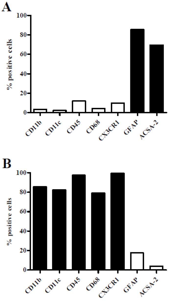
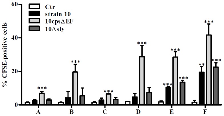
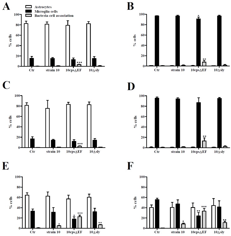
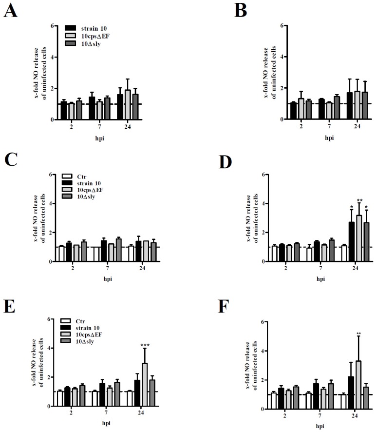
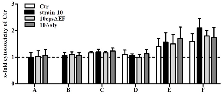
Streptococcus (S.) suis infections are the most common cause of meningitis in pigs. Moreover, S. suis is a zoonotic pathogen, which can lead to meningitis in humans, mainly in adults. We assume that glial cells may play a crucial role in host-pathogen interactions during S. suis infection of the central nervous system. Glial cells are considered to possess important functions during inflammation and injury of the brain in bacterial meningitis. In the present study, we established primary astrocyte-microglial cell co-cultures to investigate interactions of S. suis with glial cells. For this purpose, microglial cells and astrocytes were isolated from new-born mouse brains and characterized by flow cytometry, followed by the establishment of astrocyte and microglial cell mono-cultures as well as astrocyte-microglial cell co-cultures. In addition, we prepared microglial cell mono-cultures co-incubated with uninfected astrocyte mono-culture supernatants and astrocyte mono-cultures co-incubated with uninfected microglial cell mono-culture supernatants. After infection of the different cell cultures with S. suis, bacteria-cell association was mainly observed with microglial cells and most prominently with a non-encapsulated mutant of S. suis. A time-dependent induction of NO release was found only in the co-cultures and after co-incubation of microglial cells with uninfected supernatants of astrocyte mono-cultures mainly after infection with the capsular mutant. Only moderate cytotoxic effects were found in co-cultured glial cells after infection with S. suis. Taken together, astrocytes and astrocyte supernatants increased interaction of microglial cells with S. suis. Astrocyte-microglial cell co-cultures are suitable to study S. suis infections and bacteria-cell association as well as NO release by microglial cells was enhanced in the presence of astrocytes.

Keywords: NO release; S. suis; astrocytes; bacteria-cell-association; co-cultures; microglial cells.


Figures

Figure 1
Figure 1
Phenotypical characterisation of primary mouse astrocytes and microglial cells. Flow cytometry analysis of primary astrocytes (A) and microglial cells (B) isolated from brain tissue of new-born C57BL/6 mice. Cells were stained for cell-surface antigens (CD11b, CD11c, CD45, CX3CR1, and ACSA-2) and intracellular antigens (CD68 and GFAP) as indicated. Means of two independent preparations are shown.
Figure 2
Figure 2
Association of S. suis with primary mouse glial cells. Various glial cell culture systems: (A) astrocyte mono-culture, (B) microglial cell mono-culture, (C) astrocyte mono-culture pre-incubated with supernatants (SN) of uninfected microglial cell cultures, (D) microglial cell mono-culture pre-incubated with SN of uninfected astrocyte cultures, (E) astrocyte-microglial cell co-culture (low amount of microglial cells), and (F) astrocyte-microglial cell co-culture (high amount of microglial cells), respectively, were infected with CFSE-labeled S. suis strain 10, 10cpsΔEF, or 10Δsly at a MOI of 10:1. Percentage of CFSE-positive cells were measured by flow cytometry. Results are expressed as means with standard deviation (SD) of three independent experiments, and statistically significant differences when compared to uninfected control cells are indicated by ** (p-value < 0.01), and *** (p-value < 0.001), one-way analysis of variance (ANOVA) followed by a Dunnett post-hoc test.
Figure 3
Figure 3
Association of S. suis with primary mouse astrocytes and microglial cells. Various glial cell culture systems: (A) astrocyte mono-culture; (B) microglial cell mono-culture; (C) astrocyte mono-culture pre-stimulated with SN of uninfected microglial cell cultures; (D) microglial cell mono-culture pre-stimulated with SN of uninfected astrocyte cultures; (E) astrocyte-microglial cell co-culture (low amount of microglial cells); and (F) astrocyte-microglial cell co-culture (high amount of microglial cells), were infected with CFSE-labeled S. suis strain 10, 10cpsΔEF, or 10Δsly at a MOI 10:1 for 2 h. Astrocytes and microglial cells were stained for the cell-surface antigens ACSA-2 and CX3CR1, respectively. Relative amounts (%) of astrocytes (white bars), microglial cells (black bars) and microglial cells/astrocytes in association with bacteria (CFSE-positive, grey bars) were determined by flow cytometry analysis. Results are expressed as means with SD of three independent experiments, and statistically significant differences when compared to uninfected control cells are indicated by * (p-value < 0.05), ** (p-value < 0.01), and *** (p-value < 0.001), one-way-ANOVA followed by a Dunnett post-hoc test.
Figure 4
Figure 4
NO release by primary mouse glial cell cultures after infection with S. suis. Various glial cell culture systems: (A) astrocyte mono-culture; (B) microglial cell mono-culture; (C) astrocyte mono-culture pre-stimulated with SN of uninfected microglial cell cultures; (D) microglial cell mono-culture pre-stimulated with SN of uninfected astrocyte cultures; (E) astrocyte-microglial cell co-culture (low amount of microglial cells); and (F) astrocyte-microglial cell co-culture (high amount of microglial cells), were infected with S. suis strain 10, 10cpsΔEF, or 10Δsly at a MOI of 10:1 for 2, 7, and 24 h (hpi). Nitirc oxide (NO) release was measured using Griess reagent. All results are expressed as x-fold NO release normalized to uninfected astrocytes or microglial cells which were not pre-incubated with SN (dashed line). In the co-culture system values were normalized to uninfected astrocytes. Uninfected astrocytes or microglial cells pre-incubated with SN only or co-cultured served as further controls (Ctr, CF). Results are expressed as means with SD of four (A + B) or six (CF) independent experiments and statistically significant differences when compared to uninfected astrocytes and microglial cells (A,B), uninfected astrocytes and microglial cells pre-incubated with supernatants (C,D) or astrocyte-microglial cell co-cultures (E,F) are indicated by * (p-value < 0.05), ** (p-value < 0.01), and *** (p-value < 0.001), one-way-ANOVA followed by Dunnett post-hoc test.
Figure 5
Figure 5
Cell viability of primary mouse glial cell cultures after infection with S. suis. Various glial cell culture systems: (A) astrocyte mono-culture; (B) microglial cell mono-culture; (C) astrocyte mono-culture pre-stimulated with SN of uninfected microglial cell cultures; (D) microglial cell mono-culture pre-stimulated with SN of uninfected astrocyte cultures; (E) astrocyte-microglial cell co-culture (low amount of microglial cells); and (F) astrocyte-microglial cell co-culture (high amount of microglial cells), were infected with S. suis strain 10, 10cpsΔEF, or 10Δsly at a MOI 10:1. 24 h post infection cell viability was determined by standard lactate dehydrogenase (LDH) release assay. Results are expressed as x-fold cytotoxicity normalized to uninfected astrocytes and microglial cells which were not pre-incubated with SN (dashed line). In the co-culture system values were normalized to uninfected astrocytes. Uninfected astrocytes or microglial cells pre-stimulated with SN only or co-cultured served as further controls (Ctr, CF). Results are expressed as means with SD of three independent experiments. No significant differences when compared to uninfected astrocytes and microglial cells (A,B), uninfected astrocytes and microglial cells pre-incubated with supernatants (C,D) or astrocyte-microglial cell co-cultures (E,F) were found, one-way-ANOVA followed by a Dunnett post-hoc test.

References

    1. Reams R.Y., Glickman L.T., Harrington D.D., Thacker H.L., Bowersock T.L. Streptococcus suis infection in swine: A retrospective study of 256 cases. Part ii. Clinical signs, gross and microscopic lesions, and coexisting microorganisms. J. Vet. Diagn. Investig. 1994;6:326–334. doi: 10.1177/104063879400600308. - DOI - PubMed
    1. Straw B.E. Diseases of Swine. 9th ed. Blackwell Publisher; Ames, IA, USA: 2006.
    1. Huong V.T., Ha N., Huy N.T., Horby P., Nghia H.D., Thiem V.D., Zhu X., Hoa N.T., Hien T.T., Zamora J., et al. Epidemiology, clinical manifestations, and outcomes of Streptococcus suis infection in humans. Emerg. Infect. Dis. 2014;20:1105–1114. doi: 10.3201/eid2007.131594. - DOI - PMC - PubMed
    1. Baums C.G., Valentin-Weigand P. Surface-associated and secreted factors of Streptococcus suis in epidemiology, pathogenesis and vaccine development. Anim. Health Res. Rev. 2009;10:65–83. doi: 10.1017/S146625230999003X. - DOI - PubMed
    1. Charland N., Harel J., Kobisch M., Lacasse S., Gottschalk M. Streptococcus suis serotype 2 mutants deficient in capsular expression. Microbiology. 1998;144:325–332. doi: 10.1099/00221287-144-2-325. - DOI - PubMed

LinkOut - more resources