

Structural Changes of Inner and Outer Choroid in Central Serous Chorioretinopathy Determined by Optical Coherence Tomography
- PMID: 27305042
- PMCID: PMC4909210
- DOI: 10.1371/journal.pone.0157190
Structural Changes of Inner and Outer Choroid in Central Serous Chorioretinopathy Determined by Optical Coherence Tomography
Abstract
Purpose: To determine the structural changes of the choroid in eyes with central serous chorioretinopathy (CSC) by enhanced depth imaging optical coherence tomography (EDI-OCT).
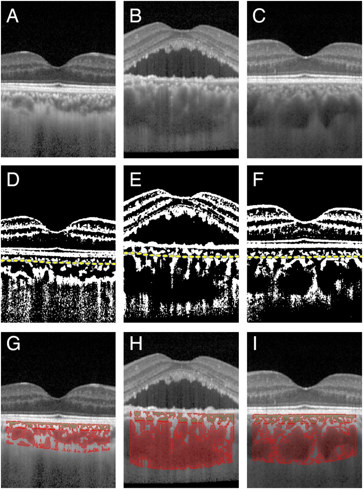
Methods: A retrospective comparative study was performed at two academic institutions. Forty eyes with CSC, their fellow eyes, and 40 eyes of age-matched controls were studied. Subfoveal cross sectional EDI-OCT images were recorded, and the hypo reflective and hyperreflective areas of the inner and outer choroid in the EDI-OCT images were separately measured. The images were analyzed by a binarization method to determine the sizes of the hyporeflective and hyperreflective areas.
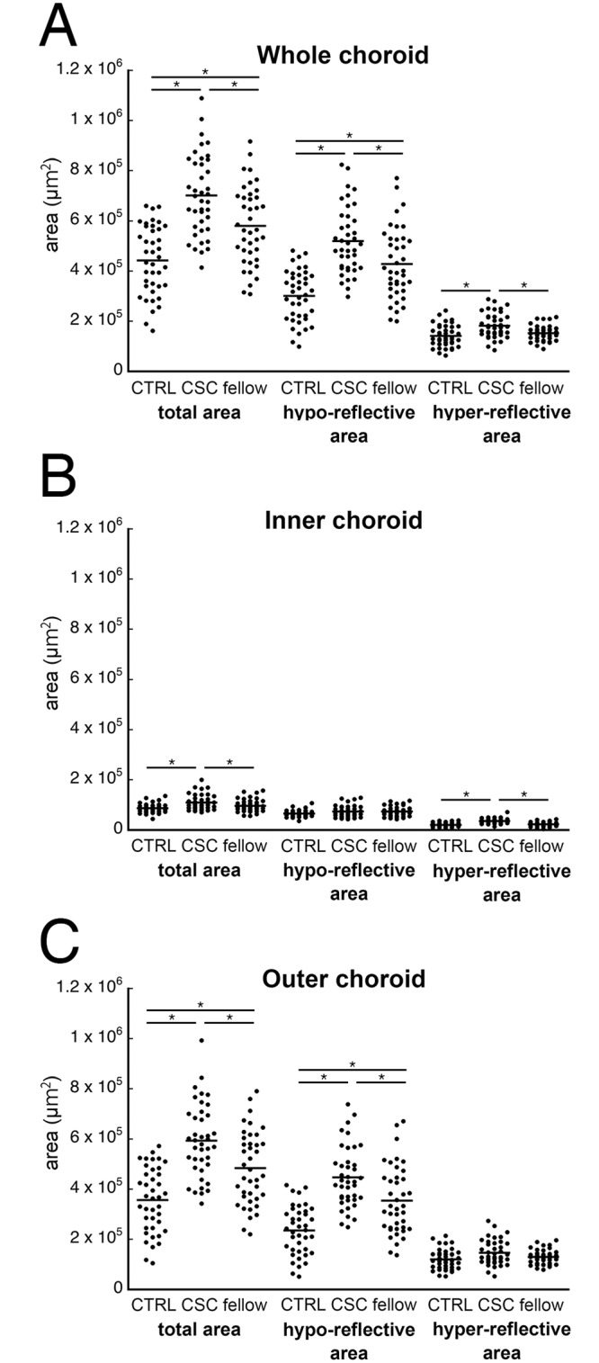
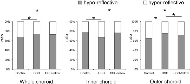
Results: In the inner choroid, the hyperreflective area was significantly larger in the CSC eyes (35,640±10,229 μm2) than the fellow eyes (22,908±8,522 μm2) and the control eyes (20,630±8,128 μm2; P<0.01 vs control for both, Wilcoxon signed-rank test). In the outer choroid, the hyporeflective area was significantly larger in the CSC eyes (446,549±121,214 μm2) than the control eyes (235,680±97,352 μm2, P<0.01). The average ratio of the hyporeflective area to the total choroidal area was smaller in the CSC eyes (67.0%) than the fellow eyes (76.5%) and the control eyes (76.7%) in the inner choroid (P<0.01, both). However, the ratio was larger in the CSC eyes (75.2%) and fellow eyes (71.7%) than in the control eyes (64.7%) in the outer choroid (P<0.01, both).
Conclusions: The larger hyperreflective area in the inner choroid is related to the inflammation and edema of the stroma of the choroid in the acute stage of CSC. The larger hyporeflective areas in the outer choroid is due to a dilatation of the vascular lumens of the larger blood vessels. These are the essential characteristics of eyes with CSC regardless of the onset.
Conflict of interest statement
Figures
References
-
- Prünte C, Flammer J. Choroidal capillary and venous congestion in central serous chorioretinopathy. Am J Ophthalmol. 1996;121:26–34. - PubMed
-
- Iida T, Kishi S, Hagimura N, Shimizu K. Persistent and bilateral choroidal vascular abnormalities in central serous chorioretinopathy. Retina. 1999;19:508–512. - PubMed
-
- Yoshioka H, Katsume Y, Akune H. Experimental central serous chorioretinopathy in monkey eyes: fluorescein angiographic findings. Ophthalmologica. 1982;185:168–178. - PubMed
Publication types
MeSH terms
Substances
LinkOut - more resources
Full Text Sources
Other Literature Sources
Medical
