

A Genetically Encoded β-Lactamase Reporter for Ultrasensitive (129) Xe NMR in Mammalian Cells
- PMID: 27305488
- PMCID: PMC5059844
- DOI: 10.1002/anie.201604055
A Genetically Encoded β-Lactamase Reporter for Ultrasensitive (129) Xe NMR in Mammalian Cells
Abstract
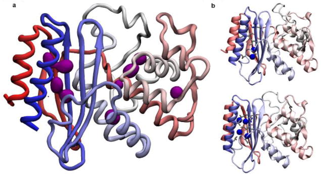
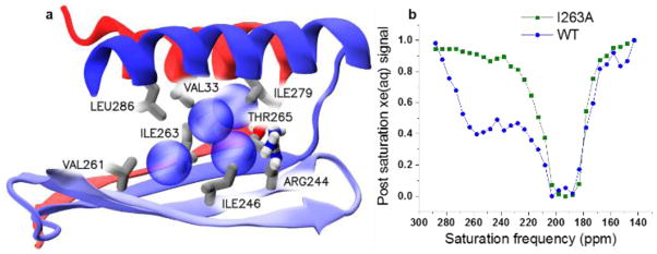
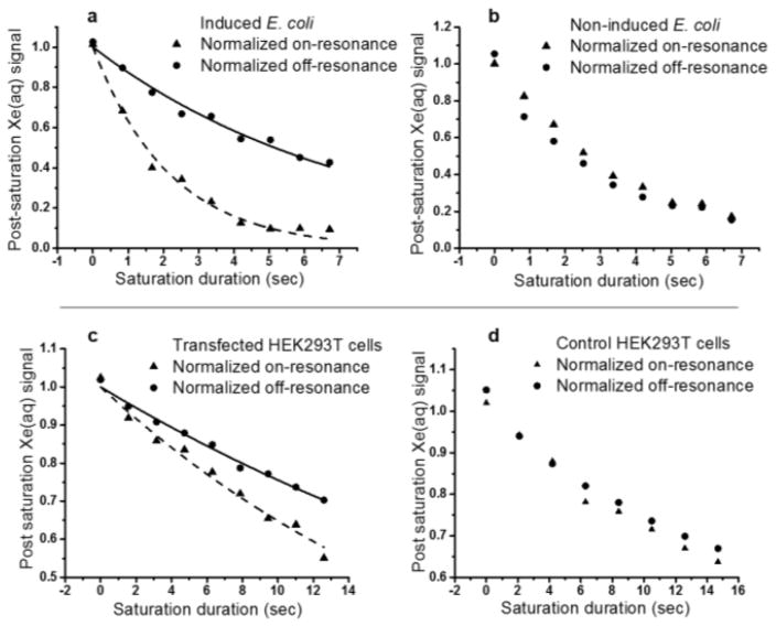
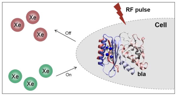
Molecular imaging holds considerable promise for elucidating biological processes in normal physiology as well as disease states, but requires noninvasive methods for identifying analytes at sub-micromolar concentrations. Particularly useful are genetically encoded, single-protein reporters that harness the power of molecular biology to visualize specific molecular processes, but such reporters have been conspicuously lacking for in vivo magnetic resonance imaging (MRI). Herein, we report TEM-1 β-lactamase (bla) as a single-protein reporter for hyperpolarized (HP) (129) Xe NMR, with significant saturation contrast at 0.1 μm. Xenon chemical exchange saturation transfer (CEST) interactions with the primary allosteric site in bla give rise to a unique saturation peak at 255 ppm, well removed (≈60 ppm downfield) from the (129) Xe-H2 O peak. Useful saturation contrast was also observed for bla expressed in bacterial cells and mammalian cells.
Keywords: NMR reporter genes; contrast agents; molecular imaging; protein biophysics; supramolecular chemistry.
© 2016 WILEY-VCH Verlag GmbH & Co. KGaA, Weinheim.
Figures
Similar articles
-
An Expanded Palette of Xenon-129 NMR Biosensors.Acc Chem Res. 2016 Oct 18;49(10):2179-2187. doi: 10.1021/acs.accounts.6b00309. Epub 2016 Sep 19. Acc Chem Res. 2016. PMID: 27643815 Free PMC article.
-
A Structural Basis for 129 Xe Hyper-CEST Signal in TEM-1 β-Lactamase.Chemphyschem. 2019 Jan 21;20(2):260-267. doi: 10.1002/cphc.201800624. Epub 2018 Sep 13. Chemphyschem. 2019. PMID: 30151973 Free PMC article.
-
Hyperpolarized Xenon-129 Chemical Exchange Saturation Transfer (HyperCEST) Molecular Imaging: Achievements and Future Challenges.Int J Mol Sci. 2024 Feb 5;25(3):1939. doi: 10.3390/ijms25031939. Int J Mol Sci. 2024. PMID: 38339217 Free PMC article. Review.
-
Nanomolar small-molecule detection using a genetically encoded 129Xe NMR contrast agent.Chem Sci. 2017 Nov 1;8(11):7631-7636. doi: 10.1039/c7sc03601a. Epub 2017 Sep 20. Chem Sci. 2017. PMID: 29568427 Free PMC article.
-
Nanoparticle-Based Contrast Agents for 129Xe HyperCEST NMR and MRI Applications.Contrast Media Mol Imaging. 2019 Nov 22;2019:9498173. doi: 10.1155/2019/9498173. eCollection 2019. Contrast Media Mol Imaging. 2019. PMID: 31819739 Free PMC article. Review.
Cited by
-
Paramagnetic Organocobalt Capsule Revealing Xenon Host-Guest Chemistry.Inorg Chem. 2020 Oct 5;59(19):13831-13844. doi: 10.1021/acs.inorgchem.9b03634. Epub 2020 Mar 24. Inorg Chem. 2020. PMID: 32207611 Free PMC article.
-
Multiplexed 129Xe HyperCEST MRI Detection of Genetically Reconstituted Bacterial Protein Nanoparticles in Human Cancer Cells.Contrast Media Mol Imaging. 2020 Mar 12;2020:5425934. doi: 10.1155/2020/5425934. eCollection 2020. Contrast Media Mol Imaging. 2020. PMID: 32256252 Free PMC article.
-
Advances in Monitoring Cell-Based Therapies with Magnetic Resonance Imaging: Future Perspectives.Int J Mol Sci. 2017 Jan 19;18(1):198. doi: 10.3390/ijms18010198. Int J Mol Sci. 2017. PMID: 28106829 Free PMC article. Review.
-
NMR Hyperpolarization Techniques of Gases.Chemistry. 2017 Jan 18;23(4):725-751. doi: 10.1002/chem.201603884. Epub 2016 Dec 5. Chemistry. 2017. PMID: 27711999 Free PMC article. Review.
-
An Expanded Palette of Xenon-129 NMR Biosensors.Acc Chem Res. 2016 Oct 18;49(10):2179-2187. doi: 10.1021/acs.accounts.6b00309. Epub 2016 Sep 19. Acc Chem Res. 2016. PMID: 27643815 Free PMC article.
References
-
- Lippincott-Schwartz J, Patterson GH. Science. 2003;300:87–91. - PubMed
-
- Walker TG, Happer W. Rev Mod Phys. 1997;69:629–642.
-
- Bai Y, Hill PA, Dmochowski IJ. Anal Chem. 2012;84:9935–9941. - PMC - PubMed
- Wang Y, Dmochowski IJ. Chem Commun. 2015;51:8982–8985. - PMC - PubMed
- Schnurr M, Sloniec-Myszk J, Dopfert J, Schroder L, Hennig A. Angew Chem Int Edit. 2015;54:13444–13447. - PubMed
- Wang Y, Roose BW, Philbin JP, Doman JL, Dmochowski IJ. Angew Chem Int Ed. 2016;55:1733–1736. - PMC - PubMed
Publication types
MeSH terms
Substances
Grants and funding
LinkOut - more resources
Full Text Sources
Other Literature Sources
Research Materials
Miscellaneous
