. 2016 Aug 1;39(8):1613-24.
doi: 10.5665/sleep.6032.

Age-Related Reduction of Recovery Sleep and Arousal Threshold in Drosophila

Affiliations

Age-Related Reduction of Recovery Sleep and Arousal Threshold in Drosophila

Julie Vienne et al. Sleep. .

Abstract

Study objectives: Physiological studies show that aging affects both sleep quality and quantity in humans, and sleep complaints increase with age. Along with knowledge about the negative effects of poor sleep on health, understanding the enigmatic relationship between sleep and aging is important. Because human sleep is similar to Drosophila (fruit fly) sleep in many ways, we addressed the effects of aging on sleep in this model organism.

Methods: Baseline sleep was recorded in five different Drosophila genotypes raised at either 21°C or 25°C. The amount of sleep recovered was then investigated after a nighttime of sleep deprivation (12 h) and after chronic sleep deprivation (3 h every night for multiple nights). Finally, the effects of aging on arousal, namely, sensitivity to neuronal and mechanical stimuli, were studied.

Results: We show that fly sleep is affected by age in a manner similar to that of humans and other mammals. Not only do older flies of several genotypes have more fragmented sleep and reduced total sleep time compared to young flies, but older flies also fail to recover as much sleep after sleep deprivation. This suggests either lower sleep homeostasis and/or a failure to properly recover sleep. Older flies also show a decreased arousal threshold, i.e., an increased response to neuronal and mechanical wake-promoting stimuli. The reduced threshold may either reflect or cause the reduced recovery sleep of older flies compared to young flies after sleep deprivation.

Conclusions: Further studies are certainly needed, but we suggest that the lower homeostatic sleep drive of older flies causes their decreased arousal threshold.

Keywords: Drosophila; aging; arousal; arousal threshold; sleep deprivation.


Figures

Figure 1
Figure 1
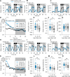
Graphical representation of how aging decreases sleep and increases sleep fragmentation under baseline conditions. A,F. Time courses of sleep (hourly values; mean ± standard error of the mean) during 24-h baseline in w1118 and Canton S flies at three different ages. (Two-way analysis of variance [ANOVA] factors Age [P < 0.0001], Time [P < 0.0001] and their interaction [P < 0.0001]; significant difference between ages: Tukey test [P < 0.05; stars]; gray areas illustrate 12-h dark periods). B,G. Total sleep duration decreased with aging in w1118 and Canton S flies in both light and dark periods. C,D,H,I. With aging during the 12-h dark period (major sleep period), the mean sleep episode duration shortened and number of sleep episodes increased, i.e., aging correlated with more fragmented sleep in both w1118 and Canton S flies. During the light period, the duration of mean sleep episodes still decreased as flies aged. However, the total number of sleep episodes varied with age and genotype. (One-way ANOVA factor Age [P < 0.003]; Tukey test [P < 0.05; star with connected lines]; for the mean sleep episode duration: Kruskal-Wallis one-way ANOVA on ranks, factor Age [P < 0.0001]; Dunn test [P < 0.05; star with connected lines]). E,J. Sleep latency was increased during the dark period in w1118 but decreased in Canton S flies (for sleep latency-light period: one-way ANOVA factor Age [P ≥ 0.22]; for sleep latency-dark period: one-way ANOVA factor Age [P = 0.0001]; Tukey test [P < 0.05; star with connected lines]). n = 166–245/age (w1118), n = 145–186/age (Canton S).
Figure 2
Figure 2
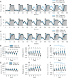
Graphical representation of how recovery sleep decreases with age after 12 h of mechanical sleep deprivation. AC, HJ. Sleep time course (hourly values; mean ± standard error of the mean) of w1118 and Canton S flies at three different ages (8, 20, and 35 days old). The 24-h baseline day was followed by a 12-h sleep deprivation (SD) during the subsequent dark period (gray area) and then a 24-h recovery period. D,K. Hourly time course of sleep gain during the 24-h recovery period after 12 h of mechanical sleep deprivation in Canton S and w1118 flies was significantly decreased with age. (Two-way analysis of variance [ANOVA], factors Age [P < 0.0001], Hour [P < 0.0001] and interaction [P < 0.0001]; Tukey test [P < 0.05; stars]). Sleep gain represents the amount of sleep gained during the recovery period compared to both baseline values and normalized by control flies (see Methods). E,F,L,M. Sleep gain and sleep recovered during the 24-h recovery period were significantly decreased with age (Kruskal-Wallis one-way ANOVA on ranks, factor Age [P < 0.001]; Dunn test [P < 0.01]; star with connected lines). Sleep recovered displays sleep gain as a percentage of sleep loss (sleep gain/sleep loss*100). G,N. Sleep loss (min) was not significantly different in w1118 but decreased in older Canton S compared to middle-aged and young flies (Kruskal-Wallis one-way ANOVA on ranks, factor Age in w1118 [P = 0.07] and in Canton S [P < 0.001]; Dunn test [P < 0.05; star with connected lines]). n = 69–108 (w1118); n = 44–82 (Canton S).
Figure 3
Figure 3
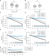
Graphical representation of how recovery sleep decreases with age after 12 h of genetic sleep deprivation. AC. Sleep time course (hourly values; mean ± standard error of the mean) of TH flies and control flies (GAL4 and UAS) at three different ages (8, 40, and 70 days old). The 24-h baseline day was followed by 12 h of sleep deprivation (SD) during the subsequent dark period (gray area) and a 24-h recovery period. D,H. Hourly time course of sleep gain during the 24-h recovery period after 12 h sleep deprivation (28°C; TH cell stimulation) in TH flies was significantly decreased with age compared to control flies (either GAL4 (D) or UAS (H). For analysis details, see Methods. For both D and H: Two-way analysis of variance [ANOVA], factor Age [P < 0.0001], Hour [P < 0.0001] and interaction [P < 0.0001]; Tukey test [P < 0.05; stars], n = 41–53. E,F,I,J. Sleep gain and sleep recovered during 24-h recovery period were significantly decreased with age (Kruskal-Wallis one-way ANOVA on ranks, factor Age [P < 0.0001]; Dunn test: [P < 0.05; stars]). Sleep recovered displays sleep gain as a percentage of sleep loss (sleep gain/sleep loss *100). G,K. Sleep loss (min) was decreased with age (Kruskal-Wallis one-way ANOVA on ranks; factor Age [P < 0.0001]; Dunn test: [P < 0.001; stars]); n = 41–52.
Figure 4
Figure 4
Graphical representation showing how young flies recover progressively more sleep after a daily 3-h sleep deprivation period compared to middle-aged flies (chronic sleep deprivation). After a baseline day (day 1), young (7 day old) and middle-aged (40 day old) TH flies were sleep-deprived during the first 3 h (black rectangles; 28°C) of the dark period (gray area) during 6 consecutive days. Between each sleep deprivation period, flies had 21 h to recover lost sleep (21°C). UAS and GAL4 flies were used as controls. A,B,C. As illustrated by the sleep time course (hourly values; mean ± standard error of the mean) of young (blue line) and middle-aged (black line) TH, UAS, and GAL4 flies, young flies recovered progressively more sleep after the daily 3 h of sleep deprivation (TH, A) than middle-aged flies. D,G. The amount of sleep loss during the 3 h of sleep deprivation varied among age and days. (vs. GAL4: two-way analysis of variance [ANOVA], factors Age [P < 0.0001], Day [P = 0.001], and interactions [P < 0.0001]; vs. UAS: two-way ANOVA, factors Age [P < 0.0001], Day [P = 0.001], and interactions [P < 0.0001]; Tukey test for factor Age [P < 0.0001; black stars]). E,F,H,I. During the 21-h recovery periods following each 3-h sleep deprivation, young TH flies gained/recovered more sleep than middle-aged TH flies (Sleep gain/recovered vs. GAL4 flies [E,F]: two-way ANOVA, factors Age [P < 0.0001], Day [P ≥ 0.4] and interactions [P ≤ 0.0003]; Sleep gain/recovered vs. UAS flies [H,I]: two-way ANOVA, factors Age [P < 0.0001], Day [P < 0.0001] and interactions [P ≤ 0.0001]; Tukey test for Age [P ≤ 0.03; black stars]). In addition, only young flies showed a progressive increase of sleep recovered with each additional 3-h sleep deprivations (H,I: Tukey test [P < 0.05; star with connected blue lines]). Sleep recovered displays sleep gain as a percentage of sleep loss (sleep gain/sleep loss *100; see also Methods for analysis details; n = 16–31 flies).
Figure 5
Figure 5
Graphical representation showing how older flies are more affected by wake-promoting stimuli than younger flies. Mild mechanical stimuli were applied to young and middle-aged Canton S flies. A,B. The percentage of flies asleep before each stimulus was higher in young flies compared to middle-aged flies, and the percentage of flies awakened by each stimulus was lower in young flies compared to middle-aged flies (one-way analysis of variance [ANOVA], factor Age [P ≤ 0.0145]; Student t-test [P < 0.0146; star with connected lines], n = 38 (young), n = 34 (middle-aged); blue dashed line in boxplots = mean, black solid line = median). During the 12-h dark period, a mild increase of temperature was applied to young, middle-aged, and old TH flies, Ecell flies, and their respective UAS and GAL4 controls. Older flies were more sensitive to this mild wake-promoting stimulus than younger flies. C,D. Cumulative difference (mean ± standard error of the mean) between the baseline dark period (21°C) and the experimental dark period (23°C) of TH flies normalized by either UAS (C) or GAL4 (D) control flies. There was an age-dependent sensitivity to wake-promoting stimuli (two-way ANOVA, factors Age [P < 0.0001], Hour [P < 0.0001], and interaction [P < 0.0001]; Tukey test [P < 0.05; stars], n = 51–55 [TH], n = 57–64 [GAL4], n = 53–62 [UAS]). E,F. Similar age-dependent sensitivity to wake-promoting stimuli was observed in Ecell flies (two-way ANOVA, factors Age [P < 0.0001], Hour [P < 0.0001] and interaction [P < 0.0001]; Tukey test [P < 0.05; stars]; n = 51–55 (Ecell), n = 57–64 (GAL4), n = 53–62 (UAS)). G. Cumulative difference between the baseline dark period and the experimental dark period of young and middle-aged Canton S flies during a 12-h subtle mechanical stimulation experiment (1 tap/10 min as described in Methods; mean ± standard error of the mean). Young fly sleep was not disrupted, whereas middle-aged flies showed a significant sleep reduction (two-way ANOVA, factors Age [P < 0.0001], Time [P < 0.0001], and interaction [P < 0.0001]; Tukey test [P < 0.05; black stars]; n = 46 (young), n = 40 (middle-aged)).

References

    1. Ingram DK, London ED, Reynolds MA. Circadian rhythmicity and sleep: effects of aging in laboratory animals. Neurobiol Aging. 1982;3:287–97. - PubMed
    1. Colas D, Cespuglio R, Sarda N. Sleep wake profile and EEG spectral power in young or old senescence accelerated mice. Neurobiol Aging. 2005;26:265–73. - PubMed
    1. Hasan S, Dauvilliers Y, Mongrain V, Franken P, Tafti M. Age-related changes in sleep in inbred mice are genotype dependent. Neurobiol Aging. 2012;33:195 e13–26. - PubMed
    1. Mendelson WB, Bergmann BM. Age-related changes in sleep in the rat. Sleep. 1999;22:145–50. - PubMed
    1. Naidoo N, Zhu J, Zhu Y, et al. Endoplasmic reticulum stress in wake-active neurons progresses with aging. Aging Cell. 2011;10:640–9. - PMC - PubMed