. 2016 Jun 16:7:11952.
doi: 10.1038/ncomms11952.

Structure of the exportin Xpo4 in complex with RanGTP and the hypusine-containing translation factor eIF5A

Affiliations

Structure of the exportin Xpo4 in complex with RanGTP and the hypusine-containing translation factor eIF5A

Metin Aksu et al. Nat Commun. .

Abstract

Xpo4 is a bidirectional nuclear transport receptor that mediates nuclear export of eIF5A and Smad3 as well as import of Sox2 and SRY. How Xpo4 recognizes such a variety of cargoes is as yet unknown. Here we present the crystal structure of the RanGTP·Xpo4·eIF5A export complex at 3.2 Å resolution. Xpo4 has a similar structure as CRM1, but the NES-binding site is occluded, and a new interaction site evolved that recognizes both globular domains of eIF5A. eIF5A contains hypusine, a unique amino acid with two positive charges, which is essential for cell viability and eIF5A function in translation. The hypusine docks into a deep, acidic pocket of Xpo4 and is thus a critical element of eIF5A's complex export signature. This further suggests that Xpo4 recognizes other cargoes differently, and illustrates how Xpo4 suppresses - in a chaperone-like manner - undesired interactions of eIF5A inside nuclei.


Figures

Figure 1
Figure 1. Improvement of Xpo4 export complex crystallization by removal of flexible parts.
(a) Export complexes comprising either full-length or truncated eIF5A were formed with Xpo4 and ZZ-bdNEDD8-tagged RanGTP. After immobilization via tagged Ran, bound proteins were eluted by bdNEDP1 protease, and analysed by SDS–polyacrylamide gel electrophoresis (SDS–PAGE) and Coomassie-staining. (b) 180 μg of the export complex was incubated with trypsin (500:1 w/w) for 90 min at 22 °C. The reaction was stopped by 5 mM PMSF and EDTA. Note that the resulting proteolyzed complex remained intact when analysed by size exclusion chromatography (SEC) on a Superdex 200 10/30 column. Experiments with chymotrypsin gave similar results. (c) Illustration of Xpo4 mutants. Protease cleavage sites were identified by mass spectrometry. (d) Overlayed size exclusion chromatograms of the export complexes derived from wild-type Xpo4 or Xpo4 loop deletions. (e) Peak fractions from d were pooled, concentrated and analysed by SDS–PAGE followed by Coomassie-staining. (f) The export complexes from e were incubated with trypsin (1,000:1 w/w) for 1 h at 22 °C and analysed by SDS–PAGE and Coomassie-staining. The ΔLoopN&C Xpo4 variant was trypsin-resistant.
Figure 2
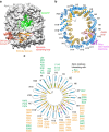
Figure 2. Structure of the RanGTP·Xpo4·eIF5A export complex and HEAT repeat organization of Xpo4.
(a) Xpo4 (grey) is shown as surface representation, while Ran (green) and eIF5A are shown as ribbon representation. Different regions of eIF5A are coloured and labelled accordingly. GTP is shown as black sticks. (b) Xpo4 in the export complex is shown in a ribbon representation (RanGTP and eIF5A are removed for clarity). Colour scheme: A helices of the HEAT repeats, blue; B helices, yellow; long inter-repeat insertions, dark pink; the acidic loop, brown. (c) Schematic representation of the Xpo4 secondary structure. Colouring is as in b. Green and orange dots represent the Xpo4 residues interacting with RanGTP and eIF5A, respectively.
Figure 3
Figure 3. RanGTP recognition by Xpo4.
The export complex is shown as a ribbon representation. On the left, Xpo4 is coloured in a gradient from blue (N-terminus) to grey (C-terminus); the acidic loop is shown in brown. eIF5A is coloured in orange and Ran in green. Switch I and II regions of Ran are shown as cyan and pink, respectively. GTP (black) is shown as sticks. The close-up on the right illustrates Xpo4-Ran interactions.
Figure 4
Figure 4. Comparison of the ligand-bound structures of CRM1 and Xpo4.
Export complexes are aligned with respect to Ran, illustrated in ribbon representations, and each shown in two different orientations (side and top views). Exportins are coloured in gradients from blue (N-terminus) to grey (C-terminus). The respective cargoes are shown in orange, Ran in green. Right, NES-binding site of CRM1 and the corresponding region of Xpo4 are shown. A and B helices of the HEAT repeats are coloured in blue and yellow, respectively. The PKI NES is coloured in orange, the NES-like fragment in Xpo4 in purple.
Figure 5
Figure 5. eIF5A recognition by Xpo4.
Xpo4 and eIF5A are rendered as surface representations, with RanGTP being removed for clarity. Rotation of eIF5A is indicated. (a) Xpo4 is coloured in grey and eIF5A in orange. Interaction surface of Xpo4 on eIF5A is shown in dark green, whereas that of eIF5A on Xpo4 in dark pink. (b) Xpo4 and eIF5A are coloured according to electrostatic potential with a colour gradient from red (negatively charged) to blue (positively charged). (c) eIF5A is coloured according to conservation with a colour gradient from cyan (variable) to maroon (conserved). Conservation was based on 50 sequences ranging from animals (human), fungi, plants to protozoans (Leishmania mexicana).
Figure 6
Figure 6. Docking of the hypusine-containing loop into the acidic pocket of Xpo4.
eIF5A is shown as orange ribbon, while the hypusine (Hpu) and histidine (H51) are shown as sticks. Xpo4 is coloured in grey and depicted as surface on the left and as ribbon on the right. The Xpo4 residues that interact with hypusine and H51 are shown as sticks. Nitrogens are shown as blue spheres, oxygens in red.
Figure 7
Figure 7. Xpo4 residues contacting hypusine and H51eIF5A are crucial for eIF5A binding and export.
(a) 1 μM Xpo4 wild type or mutants were incubated with 0.75 μM ZZ-bdNEDD8-tagged RanGTP and 1.25 μM hypusinated eIF5A in a 100 mM NaCl buffer. Formed complexes were retrieved via tagged Ran, eluted by (the tag-cleaving) bdNEDP1 protease, and analysed by SDS–polyacrylamide gel electrophoresis (SDS–PAGE) and Coomassie-staining. Uncropped gels are shown in Supplementary Fig. 5. (b) Alexa568-labelled hypusinated eIF5A (2 μM) was allowed to diffuse into the nuclei of digitonin-permeabilized HeLa cells in the presence of an energy-regenerating system and an NTR-depleted extract prepared from unfertilized Xenopus eggs. The mixture was split 15 min later and indicated Xpo4 variants (2 μM) were added. After 30 min, eIF5A distributions were recorded by confocal fluorescence microscopy. Scale bar, 20 μm. (c) The binding assay was performed as in a, but 1 μM untagged RanGTP and Xpo4 were incubated with 0.75 μM ZZ-bdSUMO tagged (non-hypusinated) wild-type or mutant eIF5A in a 50 mM NaCl buffer, and bdSENP1 was used for elution.

References

    1. Sloan K. E., Gleizes P. E. & Bohnsack M. T. Nucleocytoplasmic transport of RNAs and RNA-protein complexes. J. Mol. Biol. 428, 2040–2059 (2015). - PubMed
    1. Hurt E. & Beck M. Towards understanding nuclear pore complex architecture and dynamics in the age of integrative structural analysis. Curr. Opin. Cell Biol. 34, 31–38 (2015). - PubMed
    1. Kabachinski G. & Schwartz T. U. The nuclear pore complex--structure and function at a glance. J. Cell Sci. 128, 423–429 (2015). - PMC - PubMed
    1. Schmidt H. B. & Görlich D. Transport Selectivity of nuclear pores, phase separation, and membraneless organelles. Trends Biochem. Sci. 41, 46–61 (2016). - PubMed
    1. Görlich D. et al. A novel class of RanGTP binding proteins. J. Cell Biol. 138, 65–80 (1997). - PMC - PubMed

Publication types

MeSH terms