

Technical Limitations of the C1q Single-Antigen Bead Assay to Detect Complement Binding HLA-Specific Antibodies
- PMID: 27306532
- PMCID: PMC5457814
- DOI: 10.1097/TP.0000000000001270
Technical Limitations of the C1q Single-Antigen Bead Assay to Detect Complement Binding HLA-Specific Antibodies
Abstract
Background: Solid-phase assays to distinguish complement binding from noncomplement binding HLA-specific antibodies have been introduced, but technical limitations may compromise their interpretation. We have examined the extent to which C1q-binding to HLA-class I single-antigen beads (SAB) is influenced by denatured HLA on SAB, antibody titre, and complement interference that causes a misleading low assessment of HLA-specific antibody levels.
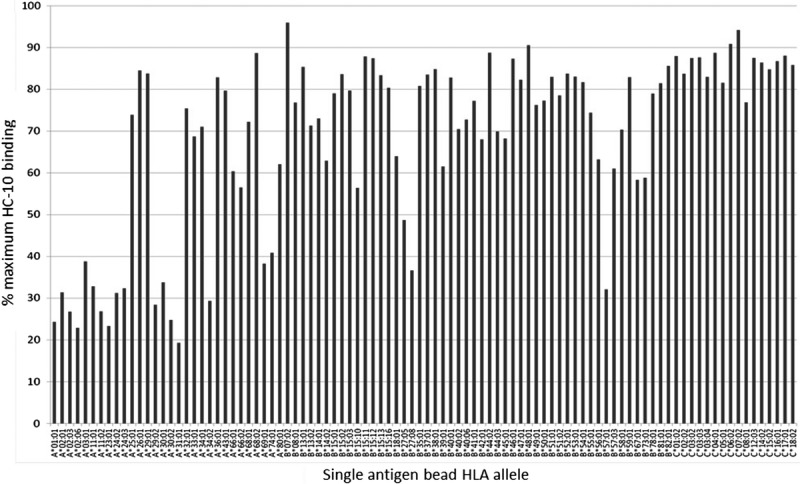
Methods: Sera from 25 highly sensitized patients were tested using Luminex IgG-SAB and C1q-SAB assays. Sera were tested undiluted, at 1:20 dilution to detect high-level IgG, and after ethylene diamine tetraacetic acid treatment to obviate complement interference. Conformational HLA and denatured HLA protein levels on SAB were determined using W6/32 and HC-10 monoclonal antibodies, respectively. Denatured HLA was expressed as HC-10 binding to untreated SAB as a percentage of maximal binding to acid-treated SAB.
Results: For undiluted sera, Luminex mean fluorescence intensity (MFI) values for IgG-SAB and C1q-SAB correlated poorly (r = 0.42). ethylene diamine tetraacetic acid and serum dilution improved the correlation (r = 0.57 and 0.77, respectively). Increasing levels of denatured HLA interfered with the detection of C1q binding. Consequently, the correlation between IgG-SAB MFI and C1q-SAB MFI was lowest using undiluted sera and SAB with greater than 30% denatured HLA (r = 0.40) and highest using diluted sera and SAB with 30% or less denatured HLA (r = 0.86).
Conclusions: Antibody level, complement interference, and denatured HLA class I on SAB may all affect the clinical interpretation of the C1q-SAB assay. The C1q-SAB assay represents a substantial additional cost for routine clinical use, and we question its justification given the potential uncertainty about its interpretation.
Conflict of interest statement
The authors declare no conflicts of interest.
Figures


Similar articles
-
Interrogating post-transplant donor HLA-specific antibody characteristics and effector functions using clinical bead assays.Hum Immunol. 2024 Nov;85(6):111094. doi: 10.1016/j.humimm.2024.111094. Epub 2024 Oct 1. Hum Immunol. 2024. PMID: 39357467
-
Comparison Between Total IgG, C1q, and C3d Single Antigen Bead Assays in Detecting Class I Complement-Binding Anti-HLA Antibodies.Transplant Proc. 2017 Nov;49(9):2031-2035. doi: 10.1016/j.transproceed.2017.09.040. Transplant Proc. 2017. PMID: 29149956
-
C1q-fixing human leukocyte antigen assay in immunized renal patients: correlation between Luminex SAB-C1q and SAB-IgG.Transplant Proc. 2012 Nov;44(9):2535-7. doi: 10.1016/j.transproceed.2012.09.084. Transplant Proc. 2012. PMID: 23146446
-
Interpreting Anti-HLA Antibody Testing Data: A Practical Guide for Physicians.Transplantation. 2016 Aug;100(8):1619-28. doi: 10.1097/TP.0000000000001203. Transplantation. 2016. PMID: 27140516 Free PMC article. Review.
-
HLA Diagnostics: Evaluating DSA Strength by Titration.Transplantation. 2018 Jan;102(1S Suppl 1):S23-S30. doi: 10.1097/TP.0000000000001817. Transplantation. 2018. PMID: 29266059 Review.
Cited by
-
EDTA Treatment for Overcoming the Prozone Effect and for Predicting C1q Binding in HLA Antibody Testing.Ann Lab Med. 2019 Nov;39(6):572-576. doi: 10.3343/alm.2019.39.6.572. Ann Lab Med. 2019. PMID: 31240886 Free PMC article.
-
Complement (C1q) Binding De Novo Donor-Specific Antibodies and Cardiac-Allograft Vasculopathy in Pediatric Heart Transplant Recipients.Transplantation. 2018 Mar;102(3):502-509. doi: 10.1097/TP.0000000000001944. Transplantation. 2018. PMID: 28885488 Free PMC article.
-
European Guideline for the Management of Kidney Transplant Patients With HLA Antibodies: By the European Society for Organ Transplantation Working Group.Transpl Int. 2022 Aug 10;35:10511. doi: 10.3389/ti.2022.10511. eCollection 2022. Transpl Int. 2022. PMID: 36033645 Free PMC article.
-
The Humoral Theory of Transplantation: Epitope Analysis and the Pathogenicity of HLA Antibodies.J Immunol Res. 2016;2016:5197396. doi: 10.1155/2016/5197396. Epub 2016 Dec 14. J Immunol Res. 2016. PMID: 28070526 Free PMC article. Review.
-
Predicting Humoral Alloimmunity from Differences in Donor and Recipient HLA Surface Electrostatic Potential.J Immunol. 2018 Dec 15;201(12):3780-3792. doi: 10.4049/jimmunol.1800683. Epub 2018 Nov 14. J Immunol. 2018. PMID: 30429288 Free PMC article.
References
-
- Gloor JM, Winters JL, Cornell LD, et al. Baseline donor-specific antibody levels and outcomes in positive crossmatch kidney transplantation. Am J Transplant. 2010;10:582–589. - PubMed
-
- Otten HG, Verhaarb MC, Borsta HPE, et al. Pretransplant donor-specific HLA class-I and -II antibodies are associated with an increased risk for kidney graft failure. Am J Transplant. 2012;12:1618–1623. - PubMed
-
- Pei R, Lee JH, Shih NJ, et al. Single human leukocyte antigen flow cytometry beads for accurate identification of human leukocyte antigen antibody specificities. Transplantation. 2003;75:43–49. - PubMed
-
- Vlad G, Ho EK, Vasilescu ER, et al. Relevance of different antibody detection methods for the prediction of antibody-mediated rejection and deceased-donor kidney allograft survival. Hum Immunol. 2009;70:589–594. - PubMed
Publication types
MeSH terms
Substances
Grants and funding
LinkOut - more resources
Full Text Sources
Other Literature Sources
Molecular Biology Databases
Research Materials
