. 2017 Apr;37(4):1374-1385.
doi: 10.1177/0271678X16655550. Epub 2016 Jan 1.

Paravascular channels, cisterns, and the subarachnoid space in the rat brain: A single compartment with preferential pathways

Affiliations

Paravascular channels, cisterns, and the subarachnoid space in the rat brain: A single compartment with preferential pathways

Beatrice Bedussi et al. J Cereb Blood Flow Metab. 2017 Apr.

Abstract

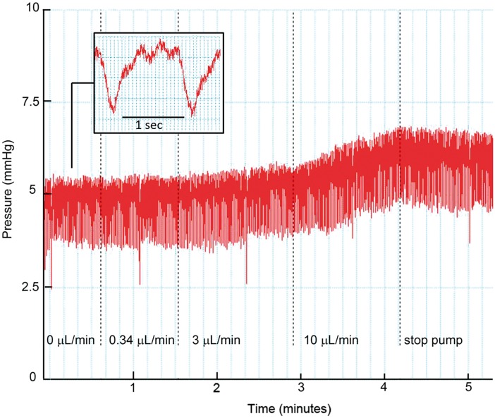
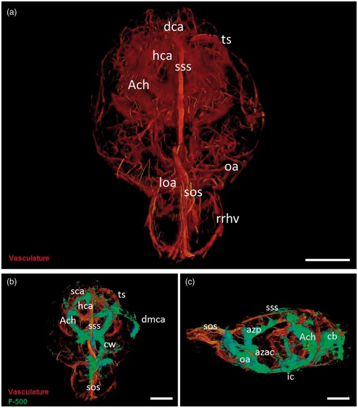
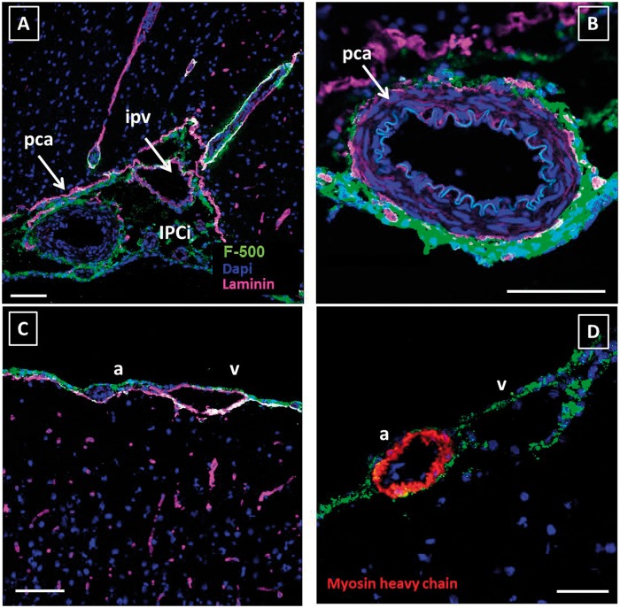
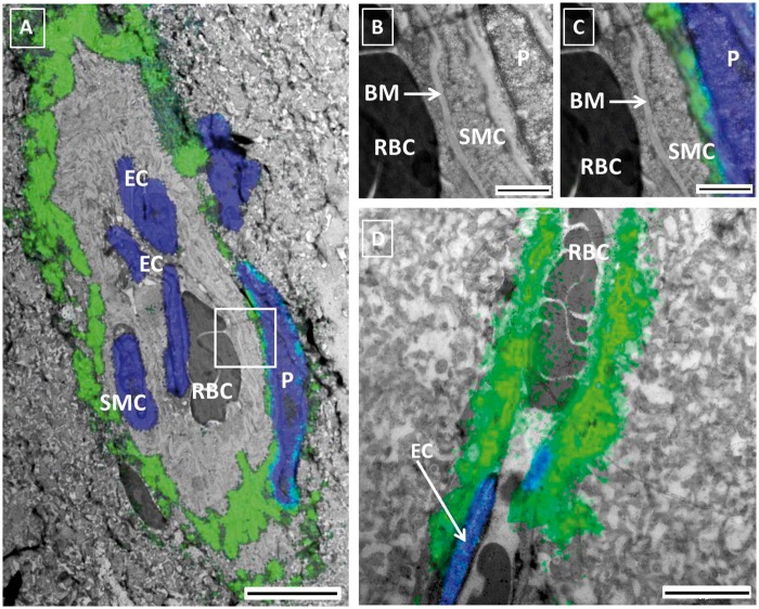
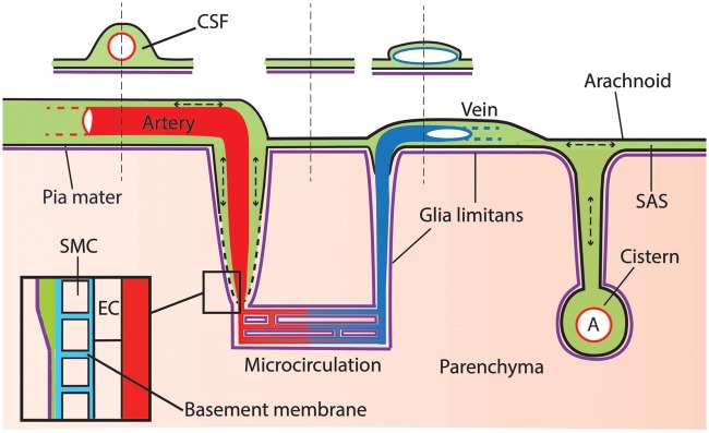
Recent evidence suggests an extensive exchange of fluid and solutes between the subarachnoid space and the brain interstitium, involving preferential pathways along blood vessels. We studied the anatomical relations between brain vasculature, cerebrospinal fluid compartments, and paravascular spaces in male Wistar rats. A fluorescent tracer was infused into the cisterna magna, without affecting intracranial pressure. Tracer distribution was analyzed using a 3D imaging cryomicrotome, confocal microscopy, and correlative light and electron microscopy. We found a strong 3D colocalization of tracer with major arteries and veins in the subarachnoid space and large cisterns, attributed to relatively large subarachnoid space volumes around the vessels. Confocal imaging confirmed this colocalization and also revealed novel cisternal connections between the subarachnoid space and ventricles. Unlike the vessels in the subarachnoid space, penetrating arteries but not veins were surrounded by tracer. Correlative light and electron microscopy images indicated that this paravascular space was located outside of the endothelial layer in capillaries and just outside of the smooth muscle cells in arteries. In conclusion, the cerebrospinal fluid compartment, consisting of the subarachnoid space, cisterns, ventricles, and para-arteriolar spaces, forms a continuous and extensive network that surrounds and penetrates the rat brain, in which mixing may facilitate exchange between interstitial fluid and cerebrospinal fluid.

Keywords: Cerebrospinal fluid; glymphatic pathway; interstitial fluid; paravascular space; subarachnoid space.


Figures

Figure 1.
Figure 1.
Changes in ICP during infusion of aCSF in the cisterna magna. Pressure (mmHg) as a function of time (min). The dotted lines indicate a stepwise increase in infusion rate (µl/min). At baseline (0 µl/min), the mean ICP was 4.8 mmHg in this example. An infusion rate of 0.34 µl/min did not change the ICP, while a further stepwise increase in infusion rate to 10 µl/min clearly raised ICP. Insert: the pressure profile was affected by the heartbeat, but more importantly, by respiration. Three heartbeats are visible between two large pressure drops induced by inspiration.
Figure 2.
Figure 2.
3D visualization of the rat cerebral vasculature and CSF tracer distribution after infusion in the cisterna magna. Panel A: 3D reconstruction of the rat brain vasculature. Vessels were filled with fluorescent cast material (orange). Panel B: Frontal view of the rat brain. Tracer (F500, green) is visible around the circle of Willis (cw), and the anterior choroidal arteries (ach); along the dorsal portion of the middle cerebral artery (dmca), in the region of the hippocampal arteries (hca), and along the major sinuses-transverse sinus (ts) superior sagittal sinus (sss), and superior olfactory sinus (sos). Panel C: Lateral view of the rat brain. Tracer is present around the azygos anterior cerebral artery (azac) that dorsally becomes the azygos pericallosal artey (azp). Signal was present also caudally along the vasculature of the cerebellum (cb) and the around the superior sagittal sinus in the SAS. Scale bar represents 5 mm.
Figure 3.
Figure 3.
Tracer distribution over the SAS and cisterns of the rat brain. Sections were stained for laminin (pink) and cell nuclei (blue). Tracer is shown in green. Panel A: Coronal section of a rat brain at approximately 5.6 mm posterior to the bregma showing some of the major cerebral cisterns. In the left upper corner a miniature of the whole section. Tracer was present along the cleft (*) between the hippocampus and the medial geniculate nucleus (MG). This space connected the interpeduncular cistern (IPCi) at the base of the brain, with the ambient cistern (ACi) and the quadrigeminal cistern (QCi). Arteries and veins followed the outline of these cisterns and were embedded in tracer. Panels B and C: horizontal sections of the rat brain at approximately −7 mm from the bregma. The stars show the presence of an extension of the interpeduncular cistern that connects the SAS with the lateral ventricle (LV). The tracer was observed along this tract, which followed the outline of the hippocampus (Hi) up to the lateral ventricle. Scale bar represents 1 mm.
Figure 4.
Figure 4.
Anatomical details of tracer distribution around blood vessels. Sections were stained for laminin (pink) and cell nuclei (blue). Tracer is shown in green. Panel A shows the interpeduncular cistern (IPCi) filled with CSF, which embedded the posterior cerebral artery (pca) and the intrapeduncular vein (ipv) Panel B: close-up of the posterior cerebral artery. Panel C: Tracer surrounded a leptomeningeal artery (a) and vein (v) but did penetrate along vessels into the parenchyma in this area. Panel D: Myosin heavy chain staining was used to discriminate arteries from veins. Both artery and vein in the SAS showed tracer signal. In all pictures, the tracer is present around the leptomeningeal vessels, in the SAS, and cisterns. Scale bar in panels A–C represents 100 µm, panel D 50 µm.
Figure 5.
Figure 5.
Paravascular distribution of CSF tracer within the brain parenchyma. In contrast to the cortex, tracer was found to penetrate into the parenchyma along vessels on the ventral side of the brain. Sections were stained for laminin (panel A; pink) or myosin heavy chain (panel B; red) and cell nuclei (blue). Tracer is shown in green. Panels A, B: adjacent slides from the same specimen. Panel A was stained for laminin, while panel B was stained for myosin heavy chain, to discriminate arteries (a) from veins (v). This revealed that tracer signal was confined to arteries only. Panels C and D show a longitudinal section of a small artery. The tracer is present around the artery and is located just outside the SMC layer. EC: endothelial cell; SMC: smooth muscle cell. Panels A and B: scale bar 100 µm; Panels C and D: scale bar 10 µm.
Figure 6.
Figure 6.
Correlative light Electron Microscopy (CLEM) of the PVS. Panel A: CLEM image of a small artery obtained after merging fluorescent (green tracer, blue nuclei) and EM images of two consecutive slices of the same brain. Red blood cells (RBC) are present in the lumen. Endothelial cells (EC) and smooth muscle cells (SMC) were identified based on location and morphology. The tracer was found in the extracellular space immediately surrounding the smooth muscle layer of the artery and in between the latter and the pericyte (P). Panels B and C are higher magnifications of the vessel wall, without the fluorescent (B) and with the fluorescent (C) signal superimposed. The basement membrane between the EC and the SMC did not present signal. Panel D shows a capillary with tracer immediately below the endothelium. Panel A: scale bar 5 µm Panels B, C: scale bar 1 µm; Panel D: scale bar 5 µm.
Figure 7.
Figure 7.
Schematic representation of the CSF compartment. Tracer injected in the cisterna magna distributes via the CSF and reaches the SAS and paravascular space, which extents along arteries into the parenchyma. Leptomeningeal vessels are bathed by the CSF. However, the space around artery and vein may be different due to the particular shape of these vessels: bigger around the circular arteries and smaller around the flattened veins (see sketched cross-section). Cerebral cisterns connect to the subarachnoid space and embed major branches of leptomeningeal arteries. Pressure oscillations produced by respiration and heart beat cause mixing within the CSF compartment. Insert: at the level of the microcirculation, the PVS disappears and fuses with the basement membrane of smooth muscle cells, pericytes, and endothelial cells. Green: CSF; red: artery; blue: vein; purple: glia limitans; light blue: basement membrane; pink: parenchyma.

References

    1. Louveau A, Smirnov I, Keyes TJ, et al. Structural and functional features of central nervous system lymphatic vessels. Nature 2015; 523: 337–341. - PMC - PubMed
    1. Aspelund A, Antila S, Proulx ST, et al. A dural lymphatic vascular system that drains brain interstitial fluid and macromolecules. J Exp Med 2015; 212: 991–999. - PMC - PubMed
    1. Iliff JJ, Wang M, Liao Y, et al. A paravascular pathway facilitates CSF flow through the brain parenchyma and the clearance of interstitial solutes, including amyloid beta. Sci Transl Med 2012; 4: 147ra11. - PMC - PubMed
    1. Carare RO, Bernardes-Silva M, Newman TA, et al. Solutes, but not cells, drain from the brain parenchyma along basement membranes of capillaries and arteries: significance for cerebral amyloid angiopathy and neuroimmunology. Neuropathol Appl Neurobiol 2008; 34: 131–144. - PubMed
    1. Hladky SB, Barrand MA. Mechanisms of fluid movement into, through and out of the brain: evaluation of the evidence. Fluids Barriers CNS 2014; 11: 26. - PMC - PubMed

MeSH terms