Review
. 2016 Aug;138 Suppl 1(Suppl Suppl 1):71-94.
doi: 10.1111/jnc.13600. Epub 2016 Jun 15.

Tau physiology and pathomechanisms in frontotemporal lobar degeneration

Affiliations
Review

Tau physiology and pathomechanisms in frontotemporal lobar degeneration

Liviu-Gabriel Bodea et al. J Neurochem. 2016 Aug.

Abstract

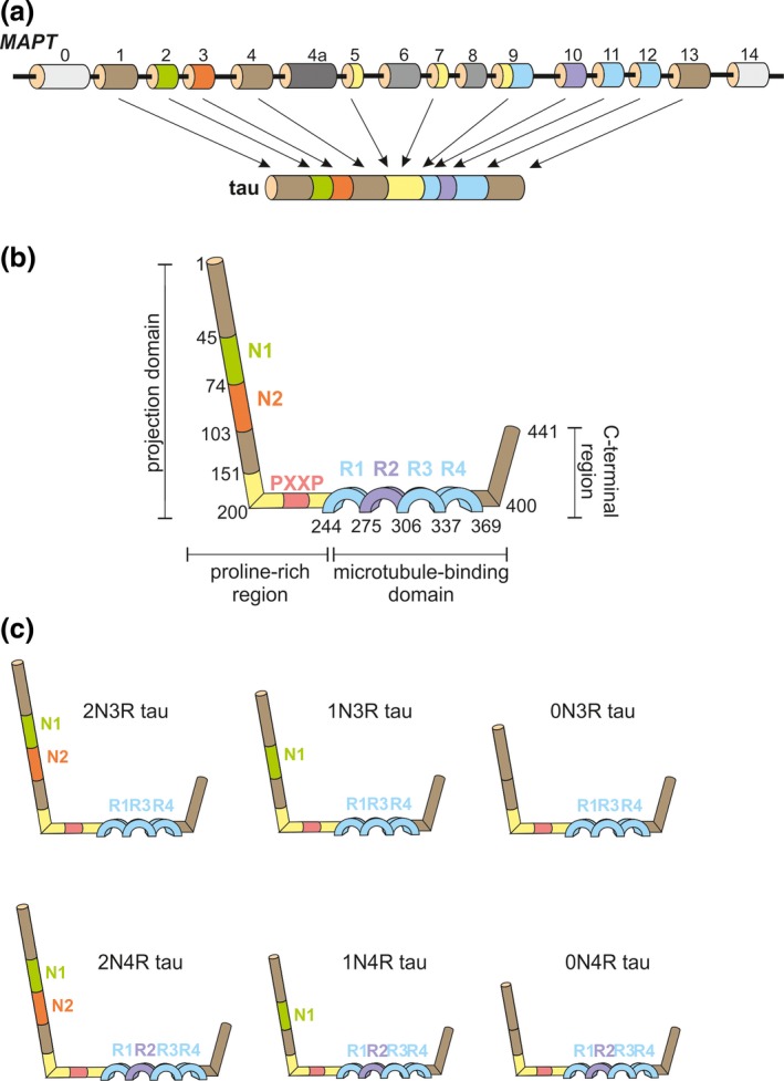
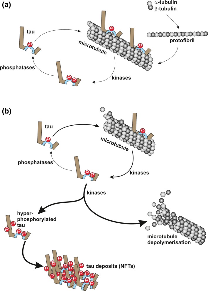
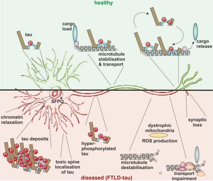
Frontotemporal lobar degeneration (FTLD) has been associated with toxic intracellular aggregates of hyperphosphorylated tau (FTLD-tau). Moreover, genetic studies identified mutations in the MAPT gene encoding tau in familial cases of the disease. In this review, we cover a range of aspects of tau function, both in the healthy and diseased brain, discussing several in vitro and in vivo models. Tau structure and function in the healthy brain is presented, accentuating its distinct compartmentalization in neurons and its role in microtubule stabilization and axonal transport. Furthermore, tau-driven pathology is discussed, introducing current concepts and the underlying experimental evidence. Different aspects of pathological tau phosphorylation, the protein's genomic and domain organization as well as its spreading in disease, together with MAPT-associated mutations and their respective models are presented. Dysfunction related to other post-transcriptional modifications and their effect on normal neuronal functions such as cell cycle, epigenetics and synapse dynamics are also discussed, providing a mechanistic explanation for the observations made in FTLD-tau cases, with the possibility for therapeutic intervention. In this review, we cover aspects of tau function, both in the healthy and diseased brain, referring to different in vitro and in vivo models. In healthy neurons, tau is compartmentalized, with higher concentrations found in the distal part of the axon. Cargo molecules are sensitive to this gradient. A disturbed tau distribution, as found in frontotemporal lobar degeneration (FTLD-tau), has severe consequences for cellular physiology: tau accumulates in the neuronal soma and dendrites, leading among others to microtubule depolymerization and impaired axonal transport. Tau forms insoluble aggregates that sequester additional molecules stalling cellular physiology. Neuronal communication is gradually lost as toxic tau accumulates in dendritic spines with subsequent degeneration of synapses and synaptic loss. Thus, by providing a mechanistic explanation for the observations made in FTLD-tau cases, arises a possibility for therapeutic interventions. This article is part of the Frontotemporal Dementia special issue.

Keywords: microtubule; phosphorylation; post-translational; spreading; synapse; transgenic.


Figures

Figure 1
Figure 1
Structure of human microtubule‐associated protein tau gene (MAPT) and protein. (a) MAPT contains 16 exons. In the brain, 11 of these exons are alternatively spliced, leading to the expression of different tau isoforms. Exons 4a, 6 and 8 are insufficiently studied. The exons are colour‐coded matching the corresponding tau protein domains; (b) The general structure of the longest human tau protein encompasses an N‐terminal projection domain, a proline‐rich region, a microtubule‐binding domain and a C‐terminal region. The N‐terminal region can contain acidic regions (N1, N2), followed by a proline‐rich region bearing up to seven PXXP motifs that can interact with other proteins via Src‐homology 3 (SH3) domains. Preceding the C‐terminal region, tau possess a microtubule‐binding domain composed of tubulin‐binding repetitions (R1, R2, R3, R4); (c) The principal 6 isoforms of human tau that are expressed in brain contain a common backbone represented by three tubulin‐binding repeats (R1, R3 and R4), to which either N1, N1 together with N2 or none of these acidic domains are added, resulting in the 2N3R, 1N3R or 0N3R isoforms. Inclusion of exon 10 of MAPT leads to the presence of the R2 tubulin‐binding domain, resulting in the 2N4R, 1N4R or 0N4R tau isoforms.
Figure 2
Figure 2
Tau‐microtubule dynamics under healthy and pathological conditions; (a) Heterodimers of α‐ and β‐tubulin assemble into protofibrils, which then form microtubules. Tau can interact with microtubules in a physiologically phosphorylated form. Increased phosphorylation of tau (e.g. as a consequence of glycogen synthase‐3, GSK3, activity) results in detachment from microtubules. Tau is reintroduced in the cycle by the action of protein phosphatases (e.g. PP2A); (b) Hyperphosphorylation of tau sequesters it from its physiological cycle (thicker arrow), resulting in the formation of intracellular tau deposits and eventually neurofibrillary tangles (NFTs), and an increasing microtubule break‐down (thickest arrows).
Figure 3
Figure 3
Tau‐mediated cellular functions under healthy and pathological conditions. In healthy neurons (top), tau molecules are found in a specific localization, with the highest concentrations being found within the distal part of the axon. Cargo molecules are particularly sensitive to the presence of a tau gradient within the axon. Disequilibrium of tau distribution (bottom), as found in frontotemporal lobar degeneration (FTLD)‐tau, has severe consequence for cellular physiology. Under these conditions, tau accumulates in the neuronal soma and dendrites, leading to microtubule depolymerization and affecting axonal transport. Mitochondrial impairments increase the production of toxic reactive oxygen species (ROS). Intra‐cytoplasmatic hyperphosphorylated tau aggregates are sequestrating more molecules, stalling cellular physiology. In the nucleus, the ratio between eu‐ and hetero‐chromatin is altered and transcription factors [e.g. splicing factor proline/glutamine‐rich (SFPQ)] can redistribute from the nucleus to the cytoplasm. Neuronal interconnectivity is lost as a result of a toxic accumulation of tau in dendritic spines, together with the loss of synaptic input.

References

    1. Ahmed Z., Bigio E. H., Budka H. et al (2013) Globular glial tauopathies (GGT): consensus recommendations. Acta Neuropathol. 126, 537–544. - PMC - PubMed
    1. Alavi Naini S. M. and Soussi‐Yanicostas N. (2015) Tau hyperphosphorylation and oxidative stress, a critical vicious circle in neurodegenerative tauopathies?. Oxid. Med. Cell. Longev. 2015, 151979. - PMC - PubMed
    1. Allen B., Ingram E., Takao M. et al (2002) Abundant tau filaments and nonapoptotic neurodegeneration in transgenic mice expressing human P301S tau protein. J. Neurosci. 22, 9340–9351. - PMC - PubMed
    1. Alonso A. C., Grundke‐Iqbal I. and Iqbal K. (1996) Alzheimer's disease hyperphosphorylated tau sequesters normal tau into tangles of filaments and disassembles microtubules. Nat. Med. 2, 783–787. - PubMed
    1. Alzheimer A. (1907) Über eine eigenartige Erkrankung der Hirnrinde. Allg. Z Psychiat. 64, 146–148.

Publication types

MeSH terms