Review
. 2016 Sep;95(10):1124-31.
doi: 10.1177/0022034516653604. Epub 2016 Jun 15.

Microglial Signaling in Chronic Pain with a Special Focus on Caspase 6, p38 MAP Kinase, and Sex Dependence

Affiliations
Review

Microglial Signaling in Chronic Pain with a Special Focus on Caspase 6, p38 MAP Kinase, and Sex Dependence

T Berta et al. J Dent Res. 2016 Sep.

Abstract

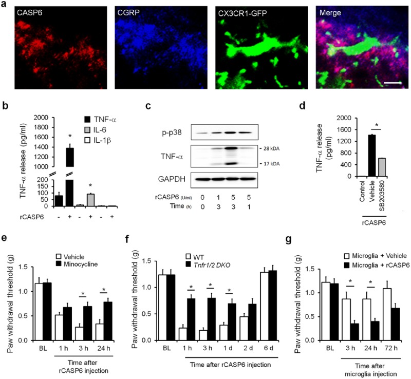
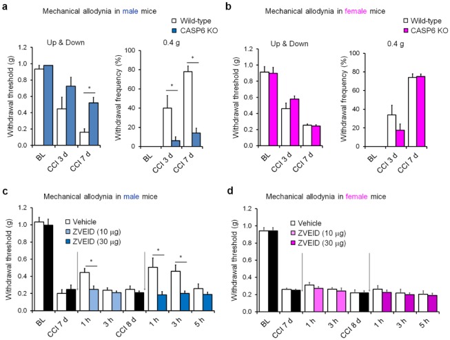
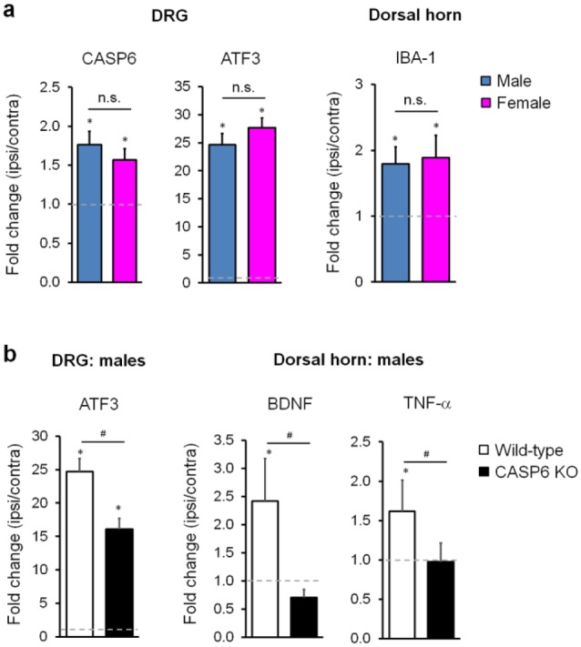
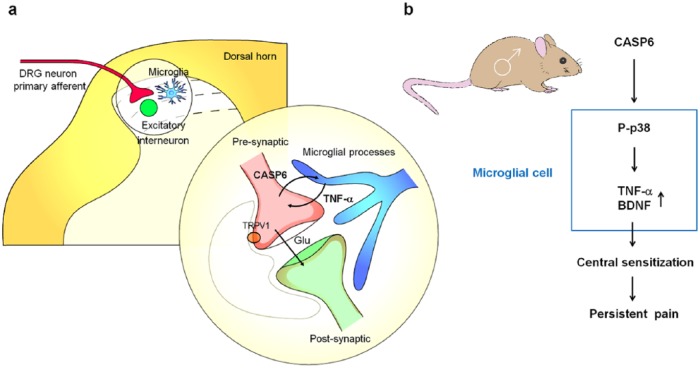
Microglia are the resident immune cells in the spinal cord and brain. Mounting evidence suggests that activation of microglia plays an important role in the pathogenesis of chronic pain, including chronic orofacial pain. In particular, microglia contribute to the transition from acute pain to chronic pain, as inhibition of microglial signaling reduces pathologic pain after inflammation, nerve injury, and cancer but not baseline pain. As compared with inflammation, nerve injury induces much more robust morphologic activation of microglia, termed microgliosis, as shown by increased expression of microglial markers, such as CD11b and IBA1. However, microglial signaling inhibitors effectively reduce inflammatory pain and neuropathic pain, arguing against the importance of morphologic activation of microglia in chronic pain sensitization. Importantly, microglia enhance pain states via secretion of proinflammatory and pronociceptive mediators, such as tumor necrosis factor α, interleukins 1β and 18, and brain-derived growth factor. Mechanistically, these mediators have been shown to enhance excitatory synaptic transmission and suppress inhibitory synaptic transmission in the pain circuits. While early studies suggested a predominant role of microglia in the induction of chronic pain, further studies have supported a role of microglia in the maintenance of chronic pain. Intriguingly, recent studies show male-dominant microglial signaling in some neuropathic pain and inflammatory pain states, although both sexes show identical morphologic activation of microglia after nerve injury. In this critical review, we provide evidence to show that caspase 6-a secreted protease that is expressed in primary afferent axonal terminals surrounding microglia-is a robust activator of microglia and induces profound release of tumor necrosis factor α from microglia via activation of p38 MAP kinase. The authors also show that microglial caspase 6/p38 signaling is male dominant in some inflammatory and neuropathic pain conditions. Finally, the authors discuss the relevance of microglial signaling in chronic trigeminal and orofacial pain.

Keywords: microglia; orofacial pain; sex-related; spinal cord; trigeminal pain; tumor necrosis factor.


Conflict of interest statement

The authors declare no potential conflicts of interest with respect to the authorship and/or publication of this article.

Figures

Figure 1.
Figure 1.
Caspase 6 (CASP6) induces mechanical allodynia via spinal microglial signaling in male mice. (a) Triple staining of CASP6, CGRP, and CX3CR1 (GFP) in the ipsilateral dorsal horn. Note close contacts between CASP6/CGRP-expressing axonal terminals and microglial cell body and processes. Scale bar, 10 µm. (b–d) Recombinant CASP6 (rCASP6) induces tumor necrosis factor α (TNF-α) release in primary microglial cultures via p38 activation. (b) Release of TNF-α and interleukins 1β and 6 (IL-1β and IL-6; ELISA analysis) in microglial culture medium after stimulation of rCASP6 (5 U/mL, 3 h). *P < 0.05, compared with control, n = 4 cultures. (c) Expression of p38 phosphorylation (p-p38) and TNF-α, revealed by Western blot analysis, in microglial cultures after rCASP6 treatment (5 U/mL, 3 h). Note a robust increase in the secreted form of TNF-α (17 kDa). (d) Effects of p38 inhibitor SB203580 (50 µM) on rCASP6-induced TNF-α release in microglial cultures. *P < 0.05, compared with vehicle (1% DMSO), n = 4 cultures. (e–g) Intrathecal injection of rCASP6- or CASP6-activated microglia induces mechanical allodynia via TNF-α secretion. BL, baseline. (e) rCASP6-induced mechanical allodynia (intrathecal [i.t.], 5 U) is reduced by minocycline pretreatment (i.t., 50 µg). *P < 0.05, n = 5 mice. (f) rCASP6-induced mechanical allodynia (i.t., 5 U) is abrogated in Tnfr double-knockout (Tnfr1/2 DKO) mice. *P < 0.05, n = 7 mice. WT, wild type. (g) Spinal (i.t.) injection of rCASP6-stimulated microglia, but not control microglia, induces mechanical allodynia. *P < 0.05, n = 5 to 7 mice. Modified from Berta et al. (2014) with permission from the journal. GAPDH, glyceraldehyde 3-phosphate dehydrogenase.
Figure 2.
Figure 2.
Spinal caspase 6 (CASP6) contributes to mechanical allodynia in male mice after nerve injury. (a, b) Chronic constriction injury (CCI)–induced mechanical allodynia in wild-type and CASP6-knockout (CASP6-KO) mice of males (a) and females (b). *P < 0.05, 2-way analysis of variance, followed by post hoc Bonferroni test, n = 5 mice/sex/group. BL, baseline. (c, d) CCI-induced mechanical allodynia in wild-type mice of males (c) and females (b) before and after intrathecal injections (arrows) of CASP6 inhibitor ZVEID (10 or 30 µg). *P < 0.05, 2-way analysis of variance, followed by post hoc Bonferroni test, n = 6 mice/sex/group. ZVEID was given 7 and 8 d after CCI. All the animal procedures were approved by the Institutional Animal Care and Use Committee of Duke University. CCI was conducted as we previously demonstrated, and mechanical allodynia was tested blindly with von Frey hairs analyzed with the up-down method (Chen et al. 2015). Mechanical allodynia was also tested by 10 stimuli with a low-threshold force (0.4 g) of von Frey hair (a, b). The doses of ZVEID were based on our previous study (Berta et al. 2014).
Figure 3.
Figure 3.
Caspase 6 (CASP6) is required for nerve injury–induced tumor necrosis factor (TNF) and brain-derived growth factor (BDNF) expression in spinal cords of male mice. (a) Chronic constriction injury (CCI) induces comparable expression of CASP6 and ATF3 in dorsal root ganglia (DRG) and IBA1 expression in the spinal cord dorsal horns of male and female mice. Gene expression was revealed by real-time quantitative polymerase chain reaction as fold change (ipsilateral side [ipsi] vs. contralateral side [contra] of the same animals). *P < 0.05, ipsi vs. contra, Student’s t test, n = 4 mice/group/sex. n.s., no significance. (b) CCI-induced expression of ATF3 in DRG and BDNF and TNF-α in dorsal horns is reduced in male mice with CASP6 deficiency. *P < 0.05, ipsi vs. contra; #P < 0.05, Student’s t test, n = 4 mice/group. DRG and spinal cord dorsal horn tissues were collected 1 week after CCI, and polymerase chain reaction analysis was conducted as previously demonstrated (Chen et al. 2015; Sorge et al. 2015).
Figure 4.
Figure 4.
Schematic illustration of axon-microglia interactions and the CASP6/p38/TNF-α signaling pathway in the spinal cord dorsal horn of male rodents. CASP6 is synthesized by C-fiber neurons of DRG and uniquely localized in axonal terminals in the superficial dorsal horn. CASP6-expressing axonal terminals form synapses with lamina IIo excitatory neurons, which in turn synapse with lamina I projection neurons to form a pain circuit (Todd 2010). These CASP6-expressing axonal terminals also have close contacts with microglial cell bodies and processes. Peripheral tissue and nerve injury results in CASP6 release from axonal terminals, and the secreted CASP6 then acts on microglial cells to trigger p38 activation. Upon activation, p38 not only induces synthesis of TNF-α and BDNF in microglia but also causes release of TNF-α and BDNF, which then act on nociceptive neurons to induce central sensitization and transition from acute pain to chronic pain. TNF-α and BDNF induce pain hypersensitivity (e.g., mechanical allodynia) via regulating both excitatory and inhibitory synaptic transmission in spinal cord pain circuits (Coull et al. 2005; Kawasaki et al. 2008; Zhang et al. 2010). It remained to be tested whether this mechanism also applies to the trigeminal system and orofacial pain. Modified from Berta et al. (2014) with permission from the journal. BDNF, brain-derived growth factor; CASP6, caspase 6; DRG, dorsal root ganglia; TNF-α, tumor necrosis factor α.

References

    1. Bale TL, Epperson CN. 2015. Sex differences and stress across the lifespan. Nat Neurosci. 18(10):1413–1420. - PMC - PubMed
    1. Berta T, Park CK, Xu ZZ, Xie RG, Liu T, Lü N, Liu YC, Ji RR. 2014. Extracellular caspase-6 drives murine inflammatory pain via microglial TNF-alpha secretion. J Clin Invest. 124(3):1173–1186. - PMC - PubMed
    1. Callahan BL, Gil AS, Levesque A, Mogil JS. 2008. Modulation of mechanical and thermal nociceptive sensitivity in the laboratory mouse by behavioral state. J Pain. 9(2):174–184. - PubMed
    1. Chen G, Park CK, Xie RG, Berta T, Nedergaard M, Ji RR. 2014. Connexin-43 induces chemokine release from spinal cord astrocytes to maintain late-phase neuropathic pain in mice. Brain. 137(Pt 8):2193–2209. - PMC - PubMed
    1. Chen G, Park CK, Xie RG, Ji RR. 2015. Intrathecal bone marrow stromal cells inhibit neuropathic pain via TGF-beta secretion. J Clin Invest. 125(8):3226–3240. - PMC - PubMed

Publication types

Substances